8S3O | pdb_00008s3o

Structure of native human CD109


Experimental Data Snapshot

  • Method: ELECTRON MICROSCOPY
  • Resolution: 2.99 Å
  • Aggregation State: PARTICLE 
  • Reconstruction Method: SINGLE PARTICLE 

Starting Model: in silico
View more details

wwPDB Validation   3D Report Full Report

Currently 8S3O does not have a validation slider image.


This is version 1.1 of the entry. See complete history


Literature

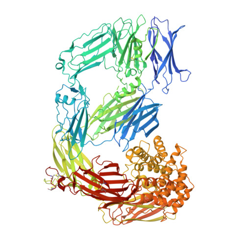
Three cryo-EM structures of CD109 reveal its mechanism of protease inhibition.

Almeida, A.V.Jensen, K.T.Harwood, S.L.Jorgensen, M.H.Nielsen, N.S.Thogersen, I.B.Enghild, J.J.Andersen, G.R.

(2025) Cell Rep 44: 115787-115787

  • DOI: https://doi.org/10.1016/j.celrep.2025.115787
  • Primary Citation of Related Structures:  
    8S3O, 9FX2, 9FX3

  • PubMed Abstract: 

    CD109 is a glycosylphosphatidylinositol-anchored protein. In addition to regulating transforming growth factor β (TGF-β) network signaling, CD109 is also a protease inhibitor. Protease cleavage of its bait region triggers a conformational change releasing the major fragment from the cell surface, exposing a reactive thioester that can conjugate proteases. To understand this protease inhibition mechanism, we determined cryoelectron microscopy structures of CD109 in native, protease-activated, and methylamine-activated conformations. Despite CD109's low sequence similarity with the protease inhibitor A2ML1, CD109 adopts a similar protease-activated conformation, suggesting a shared mechanism of protease inhibition. Deglycosylation of CD109 does not affect chymotrypsin conjugation but enhances substrate access, suggesting that CD109 glycans contribute to protease inhibition. Our data provide a structural basis for understanding CD109's protease-triggered membrane release, its protease inhibitory mechanism, and additional biological functions.


  • Organizational Affiliation
    • Department of Molecular Biology and Genetics, Aarhus University, Universitetsbyen 81, 8000 Aarhus C, Denmark.

Macromolecules
Find similar proteins by:  (by identity cutoff)  |  3D Structure
Entity ID: 1
MoleculeChains Sequence LengthOrganismDetailsImage
CD109 antigen1,447Homo sapiensMutation(s): 0 
Gene Names: CD109CPAMD7
UniProt & NIH Common Fund Data Resources
Find proteins for Q6YHK3 (Homo sapiens)
Explore Q6YHK3 
Go to UniProtKB:  Q6YHK3
PHAROS:  Q6YHK3
GTEx:  ENSG00000156535 
Entity Groups  
Sequence Clusters30% Identity50% Identity70% Identity90% Identity95% Identity100% Identity
UniProt GroupQ6YHK3
Glycosylation
Glycosylation Sites: 6Go to GlyGen: Q6YHK3-1
Sequence Annotations
Expand
  • Reference Sequence
Oligosaccharides

Help

Entity ID: 2
MoleculeChains Length2D Diagram Glycosylation3D Interactions
2-acetamido-2-deoxy-beta-D-glucopyranose-(1-4)-2-acetamido-2-deoxy-beta-D-glucopyranose
B, C
2N-Glycosylation
Glycosylation Resources
GlyTouCan:  G42666HT
GlyCosmos:  G42666HT
GlyGen:  G42666HT
Experimental Data & Validation

Experimental Data

  • Method: ELECTRON MICROSCOPY
  • Resolution: 2.99 Å
  • Aggregation State: PARTICLE 
  • Reconstruction Method: SINGLE PARTICLE 
EM Software:
TaskSoftware PackageVersion
RECONSTRUCTIONcryoSPARC4.04
MODEL REFINEMENTPHENIX1.20

Structure Validation

View Full Validation Report

Currently 8S3O does not have a validation slider image.


Entry History & Funding Information

Deposition Data


Funding OrganizationLocationGrant Number
Danish Council for Independent ResearchDenmarkDFF-2032-00111B

Revision History  (Full details and data files)

  • Version 1.0: 2025-03-05
    Type: Initial release
  • Version 1.1: 2025-09-17
    Changes: Data collection, Database references