8SZ2 | pdb_00008sz2

Stx2A1 bound to P8 stalk peptide


Experimental Data Snapshot

  • Method: X-RAY DIFFRACTION
  • Resolution: 2.01 Å
  • R-Value Free: 
    0.211 (Depositor), 0.210 (DCC) 
  • R-Value Work: 
    0.173 (Depositor), 0.170 (DCC) 
  • R-Value Observed: 
    0.175 (Depositor) 

Starting Model: experimental
View more details

wwPDB Validation   3D Report Full Report


This is version 1.2 of the entry. See complete history


Literature

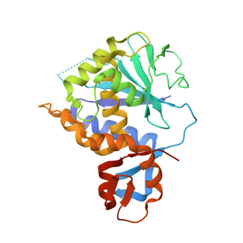
Structure-Function Analysis of the A1 Subunit of Shiga Toxin 2 with Peptides That Target the P-Stalk Binding Site and Inhibit Activity.

Li, X.P.Rudolph, M.J.Chen, Y.Tumer, N.E.

(2024) Biochemistry 63: 893-905

  • DOI: https://doi.org/10.1021/acs.biochem.3c00733
  • Primary Citation of Related Structures:  
    8SZ2

  • PubMed Abstract: 

    Shiga toxin 2a (Stx2a) is the virulence factor of Escherichia coli (STEC), which is associated with hemolytic uremic syndrome, the leading cause of pediatric kidney failure. The A1 subunit of Stx2a (Stx2A1) binds to the conserved C-terminal domain (CTD) of the ribosomal P-stalk proteins to remove an adenine from the sarcin-ricin loop (SRL) in the 28S rRNA, inhibiting protein synthesis. There are no antidotes against Stx2a or any other ribosome-inactivating protein (RIP). The structural and functional details of the binding of Stx2A1 to the P-stalk CTD are not known. Here, we carry out a deletion analysis of the conserved P-stalk CTD and show that the last eight amino acids (P8) of the P-stalk proteins are the minimal sequence required for optimal affinity and maximal inhibitory activity against Stx2A1. We determined the first X-ray crystal structure of Stx2A1 alone and in complex with P8 and identified the exact binding site. The C-terminal aspartic acid of the P-stalk CTD serves as an anchor, forming key contacts with the conserved arginine residues at the P-stalk binding pocket of Stx2A1. Although the ricin A subunit (RTA) binds to the P-stalk CTD, the last aspartic acid is more critical for the interaction with Stx2A1, indicating that RIPs differ in their requirements for the P-stalk. These results demonstrate that the catalytic activity of Stx2A1 is inhibited by blocking its interactions with the P-stalk, providing evidence that P-stalk binding is an essential first step in the recruitment of Stx2A1 to the SRL for depurination.


  • Organizational Affiliation
    • Department of Plant Biology, Rutgers, The State University of New Jersey, 59 Dudley Road, New Brunswick, New Jersey 08901, United States.

Macromolecules
Find similar proteins by:  (by identity cutoff)  |  3D Structure
Entity ID: 1
MoleculeChains Sequence LengthOrganismDetailsImage
Shiga-like toxin 2 subunit A
A, B
251Escherichia coli O104:H4Mutation(s): 0 
Gene Names: stxA2stx2AL0103
EC: 3.2.2.22
UniProt
Find proteins for P09385 (Escherichia phage 933W)
Explore P09385 
Go to UniProtKB:  P09385
Entity Groups  
Sequence Clusters30% Identity50% Identity70% Identity90% Identity95% Identity100% Identity
UniProt GroupP09385
Sequence Annotations
Expand
  • Reference Sequence

Find similar proteins by:  Sequence   |   3D Structure  

Entity ID: 2
MoleculeChains Sequence LengthOrganismDetailsImage
P stalk peptide P8C [auth P],
D [auth Q]
6Escherichia coliMutation(s): 0 
Sequence Annotations
Expand
  • Reference Sequence
Small Molecules
Ligands 4 Unique
IDChains Name / Formula / InChI Key2D Diagram3D Interactions
2PE
Query on 2PE

Download Ideal Coordinates CCD File 
E [auth A],
F [auth A]
NONAETHYLENE GLYCOL
C18 H38 O10
YZUUTMGDONTGTN-UHFFFAOYSA-N
PO4
Query on PO4

Download Ideal Coordinates CCD File 
I [auth A],
J [auth A],
K [auth A],
N [auth B],
O [auth B]
PHOSPHATE ION
O4 P
NBIIXXVUZAFLBC-UHFFFAOYSA-K
EDO
Query on EDO

Download Ideal Coordinates CCD File 
G [auth A],
H [auth A],
M [auth B]
1,2-ETHANEDIOL
C2 H6 O2
LYCAIKOWRPUZTN-UHFFFAOYSA-N
CL
Query on CL

Download Ideal Coordinates CCD File 
L [auth A],
P [auth B],
Q [auth B]
CHLORIDE ION
Cl
VEXZGXHMUGYJMC-UHFFFAOYSA-M
Experimental Data & Validation

Experimental Data

  • Method: X-RAY DIFFRACTION
  • Resolution: 2.01 Å
  • R-Value Free:  0.211 (Depositor), 0.210 (DCC) 
  • R-Value Work:  0.173 (Depositor), 0.170 (DCC) 
  • R-Value Observed: 0.175 (Depositor) 
Space Group: P 63
Unit Cell:
Length ( Å )Angle ( ˚ )
a = 102.349α = 90
b = 102.349β = 90
c = 92.891γ = 120
Software Package:
Software NamePurpose
PHENIXrefinement
HKL-2000data scaling
HKL-2000data reduction
PHASERphasing

Structure Validation

View Full Validation Report


Entry History & Funding Information

Deposition Data


Funding OrganizationLocationGrant Number
National Institutes of Health/National Institute Of Allergy and Infectious Diseases (NIH/NIAID)United States1R01AI141635-01A1

Revision History  (Full details and data files)

  • Version 1.0: 2024-05-29
    Type: Initial release
  • Version 1.1: 2024-11-20
    Changes: Structure summary
  • Version 1.2: 2024-12-11
    Changes: Database references