8V7D | pdb_00008v7d

Human DNA polymerase eta-DNA-dT primer rCTP insertion ternary complex at pH7.0 (K+ MES) with 1 Ca2+ ion


Experimental Data Snapshot

  • Method: X-RAY DIFFRACTION
  • Resolution: 1.95 Å
  • R-Value Free: 
    0.230 (Depositor), 0.230 (DCC) 
  • R-Value Work: 
    0.182 (Depositor), 0.180 (DCC) 
  • R-Value Observed: 
    0.185 (Depositor) 

Starting Model: experimental
View more details

wwPDB Validation   3D Report Full Report


Ligand Structure Quality Assessment 


This is version 1.2 of the entry. See complete history


Literature

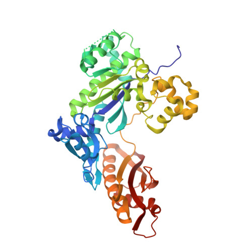
Sugar ring alignment and dynamics underline cytarabine and gemcitabine inhibition on Pol eta catalyzed DNA synthesis.

Chang, C.Zhou, G.Lee Luo, C.Eleraky, S.Moradi, M.Gao, Y.

(2024) J Biological Chem 300: 107361-107361

  • DOI: https://doi.org/10.1016/j.jbc.2024.107361
  • Primary Citation of Related Structures:  
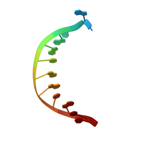
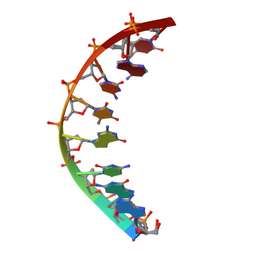
    8V7A, 8V7B, 8V7C, 8V7D, 8V7E, 8V7F, 8V7G, 8V7H, 8V7I, 8V7J, 8V7K

  • PubMed Abstract: 

    Nucleoside analogue drugs are pervasively used as antiviral and chemotherapy agents. Cytarabine and gemcitabine are anti-cancer nucleoside analogue drugs that contain C2' modifications on the sugar ring. Despite carrying all the required functional groups for DNA synthesis, these two compounds inhibit DNA extension once incorporated into DNA. It remains unclear how the C2' modifications on cytarabine and gemcitabine affect the polymerase active site during substrate binding and DNA extension. Using steady-state kinetics, static and time-resolved X-ray crystallography with DNA polymerase η (Pol η) as a model system, we showed that the sugar ring C2' chemical groups on cytarabine and gemcitabine snugly fit within the Pol η active site without occluding the steric gate. During DNA extension, Pol η can extend past gemcitabine but with much lower efficiency past cytarabine. The Pol η crystal structures show that the -OH modification in the β direction on cytarabine locks the sugar ring in an unfavorable C2'-endo geometry for product formation. On the other hand, the addition of fluorine atoms on gemcitabine alters the proper conformational transition of the sugar ring for DNA synthesis. Our study illustrates mechanistic insights into chemotherapeutic drug inhibition and resistance and guides future optimization of nucleoside analogue drugs.


  • Organizational Affiliation

    Department of Biosciences, Rice University, Houston, Texas, USA.


Macromolecules

Find similar proteins by:  (by identity cutoff)  |  3D Structure
Entity ID: 1
MoleculeChains Sequence LengthOrganismDetailsImage
DNA polymerase eta435Homo sapiensMutation(s): 0 
Gene Names: POLHRAD30RAD30AXPV
EC: 2.7.7.7
UniProt & NIH Common Fund Data Resources
Find proteins for Q9Y253 (Homo sapiens)
Explore Q9Y253 
Go to UniProtKB:  Q9Y253
PHAROS:  Q9Y253
GTEx:  ENSG00000170734 
Entity Groups  
Sequence Clusters30% Identity50% Identity70% Identity90% Identity95% Identity100% Identity
UniProt GroupQ9Y253
Sequence Annotations
Expand
  • Reference Sequence

Find similar nucleic acids by:  Sequence   |   3D Structure  

Entity ID: 2
MoleculeChains LengthOrganismImage
DNA (5'-D(*CP*AP*TP*GP*AP*TP*GP*AP*CP*GP*CP*T)-3')B [auth T]12synthetic construct
Sequence Annotations
Expand
  • Reference Sequence

Find similar nucleic acids by:  Sequence   |   3D Structure  

Entity ID: 3
MoleculeChains LengthOrganismImage
DNA (5'-D(*AP*GP*CP*GP*TP*CP*AP*T*())-3')C [auth P]8synthetic construct
Sequence Annotations
Expand
  • Reference Sequence
Small Molecules
Ligands 4 Unique
IDChains Name / Formula / InChI Key2D Diagram3D Interactions
CTP (Subject of Investigation/LOI)
Query on CTP

Download Ideal Coordinates CCD File 
G [auth A]CYTIDINE-5'-TRIPHOSPHATE
C9 H16 N3 O14 P3
PCDQPRRSZKQHHS-XVFCMESISA-N
GOL
Query on GOL

Download Ideal Coordinates CCD File 
D [auth A],
E [auth A],
F [auth A]
GLYCEROL
C3 H8 O3
PEDCQBHIVMGVHV-UHFFFAOYSA-N
CA
Query on CA

Download Ideal Coordinates CCD File 
H [auth A]CALCIUM ION
Ca
BHPQYMZQTOCNFJ-UHFFFAOYSA-N
K
Query on K

Download Ideal Coordinates CCD File 
I [auth A]POTASSIUM ION
K
NPYPAHLBTDXSSS-UHFFFAOYSA-N
Experimental Data & Validation

Experimental Data

  • Method: X-RAY DIFFRACTION
  • Resolution: 1.95 Å
  • R-Value Free:  0.230 (Depositor), 0.230 (DCC) 
  • R-Value Work:  0.182 (Depositor), 0.180 (DCC) 
  • R-Value Observed: 0.185 (Depositor) 
Space Group: P 61
Unit Cell:
Length ( Å )Angle ( ˚ )
a = 99.058α = 90
b = 99.058β = 90
c = 81.386γ = 120
Software Package:
Software NamePurpose
PHENIXrefinement
XSCALEdata scaling
XDSdata reduction
MOLREPphasing

Structure Validation

View Full Validation Report


Ligand Structure Quality Assessment 


Entry History & Funding Information

Deposition Data


Funding OrganizationLocationGrant Number
Cancer Prevention and Research Institute of Texas (CPRIT)United StatesRR190046
Welch FoundationUnited StatesC-2033-20200401
National Institutes of Health/National Institute of General Medical Sciences (NIH/NIGMS)United StatesT32 GM008280

Revision History  (Full details and data files)

  • Version 1.0: 2024-05-15
    Type: Initial release
  • Version 1.1: 2025-05-28
    Changes: Database references, Structure summary
  • Version 1.2: 2025-06-04
    Changes: Database references