8VC5 | pdb_00008vc5

Crystal structure of glutamyl-tRNA synthetase GluRS from Pseudomonas aeruginosa (Zinc bound)


Experimental Data Snapshot

  • Method: X-RAY DIFFRACTION
  • Resolution: 2.30 Å
  • R-Value Free: 
    0.256 (Depositor), 0.250 (DCC) 
  • R-Value Work: 
    0.208 (Depositor), 0.210 (DCC) 
  • R-Value Observed: 
    0.210 (Depositor) 

Starting Model: experimental
View more details

wwPDB Validation   3D Report Full Report


Ligand Structure Quality Assessment 


This is version 1.2 of the entry. See complete history


Literature

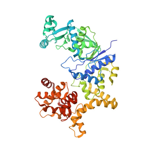
Architecture of Pseudomonas aeruginosa glutamyl-tRNA synthetase defines a subfamily of dimeric class Ib aminoacyl-tRNA synthetases.

Fenwick, M.K.Mayclin, S.J.Seibold, S.DeRocher, A.E.Subramanian, S.Phan, I.Q.Dranow, D.M.Lorimer, D.D.Abramov, A.B.Choi, R.Hewitt, S.N.Edwards, T.E.Bullard, J.M.Battaile, K.P.Wower, I.K.Soe, A.C.Tsutakawa, S.E.Lovell, S.Myler, P.J.Wower, J.Staker, B.L.

(2025) Proc Natl Acad Sci U S A 122: e2504757122-e2504757122

  • DOI: https://doi.org/10.1073/pnas.2504757122
  • Primary Citation of Related Structures:  
    8VC5

  • PubMed Abstract: 

    The aminoacyl-tRNA synthetases (AaRSs) are an ancient family of structurally diverse enzymes that are divided into two major classes. The functionalities of most AaRSs are inextricably linked to their oligomeric states. While GluRSs were previously classified as monomers, the current investigation reveals that the form expressed in Pseudomonas aeruginosa is a rotationally pseudosymmetrical homodimer featuring intersubunit tRNA binding sites. Both subunits display a highly bent, "pipe strap" conformation, with the anticodon binding domain directed toward the active site. The tRNA binding sites are similar in shape to those of the monomeric GluRSs, but are formed through an approximately 180-degree rotation of the anticodon binding domains and dimerization via the anticodon and D-arm binding domains. As a result, each anticodon binding domain is poised to recognize the anticodon loop of a tRNA bound to the adjacent protomer. Additionally, the anticodon binding domain has an α-helical C -terminal extension containing a conserved lysine-rich consensus motif positioned near the predicted location of the acceptor arm, suggesting dual functions in tRNA recognition. The unique architecture of Pa GluRS broadens the structural diversity of the GluRS family, and member synthetases of all bacterial AaRS subclasses have now been identified that exhibit oligomerization.


  • Organizational Affiliation
    • Seattle Structural Genomics Center for Infectious Disease, Seattle Children's Research Institute, Seattle, WA 98109.

Macromolecules
Find similar proteins by:  (by identity cutoff)  |  3D Structure
Entity ID: 1
MoleculeChains Sequence LengthOrganismDetailsImage
Glutamate--tRNA ligase
A, B
502Pseudomonas aeruginosa PAO1Mutation(s): 0 
Gene Names: gltXPA3134
EC: 6.1.1.17
UniProt
Find proteins for Q9XCL6 (Pseudomonas aeruginosa (strain ATCC 15692 / DSM 22644 / CIP 104116 / JCM 14847 / LMG 12228 / 1C / PRS 101 / PAO1))
Explore Q9XCL6 
Go to UniProtKB:  Q9XCL6
Entity Groups  
Sequence Clusters30% Identity50% Identity70% Identity90% Identity95% Identity100% Identity
UniProt GroupQ9XCL6
Sequence Annotations
Expand
  • Reference Sequence
Small Molecules
Ligands 5 Unique
IDChains Name / Formula / InChI Key2D Diagram3D Interactions
2PE
Query on 2PE

Download Ideal Coordinates CCD File 
G [auth A]NONAETHYLENE GLYCOL
C18 H38 O10
YZUUTMGDONTGTN-UHFFFAOYSA-N
FLC (Subject of Investigation/LOI)
Query on FLC

Download Ideal Coordinates CCD File 
C [auth A],
H [auth B]
CITRATE ANION
C6 H5 O7
KRKNYBCHXYNGOX-UHFFFAOYSA-K
SO4
Query on SO4

Download Ideal Coordinates CCD File 
D [auth A],
E [auth A],
I [auth B]
SULFATE ION
O4 S
QAOWNCQODCNURD-UHFFFAOYSA-L
GOL
Query on GOL

Download Ideal Coordinates CCD File 
J [auth B]GLYCEROL
C3 H8 O3
PEDCQBHIVMGVHV-UHFFFAOYSA-N
ZN (Subject of Investigation/LOI)
Query on ZN

Download Ideal Coordinates CCD File 
F [auth A],
K [auth B]
ZINC ION
Zn
PTFCDOFLOPIGGS-UHFFFAOYSA-N
Experimental Data & Validation

Experimental Data

  • Method: X-RAY DIFFRACTION
  • Resolution: 2.30 Å
  • R-Value Free:  0.256 (Depositor), 0.250 (DCC) 
  • R-Value Work:  0.208 (Depositor), 0.210 (DCC) 
  • R-Value Observed: 0.210 (Depositor) 
Space Group: P 21 21 21
Unit Cell:
Length ( Å )Angle ( ˚ )
a = 46.814α = 90
b = 134.833β = 90
c = 181.625γ = 90
Software Package:
Software NamePurpose
PHENIXrefinement
Aimlessdata scaling
XDSdata reduction
PHASERphasing

Structure Validation

View Full Validation Report


Ligand Structure Quality Assessment 


Entry History & Funding Information

Deposition Data


Funding OrganizationLocationGrant Number
National Institutes of Health/National Institute Of Allergy and Infectious Diseases (NIH/NIAID)United States75N93022C00036
National Institutes of Health/Office of the DirectorUnited StatesS10OD030394

Revision History  (Full details and data files)

  • Version 1.0: 2023-12-27
    Type: Initial release
  • Version 1.1: 2025-04-30
    Changes: Database references, Structure summary
  • Version 1.2: 2025-05-21
    Changes: Database references