8VVN | pdb_00008vvn

Cryo-EM structure of a type I ZorAB complex from Shewanella sp. strain ANA-3


Experimental Data Snapshot

  • Method: ELECTRON MICROSCOPY
  • Resolution: 2.20 Å
  • Aggregation State: PARTICLE 
  • Reconstruction Method: SINGLE PARTICLE 

wwPDB Validation   3D Report Full Report


This is version 1.2 of the entry. See complete history


Literature

Modularity of Zorya defense systems during phage inhibition.

Mariano, G.Deme, J.C.Readshaw, J.J.Grobbelaar, M.J.Keenan, M.El-Masri, Y.Bamford, L.Songra, S.Blower, T.R.Palmer, T.Lea, S.M.

(2025) Nat Commun 16: 2344-2344

  • DOI: https://doi.org/10.1038/s41467-025-57397-2
  • Primary Citation of Related Structures:  
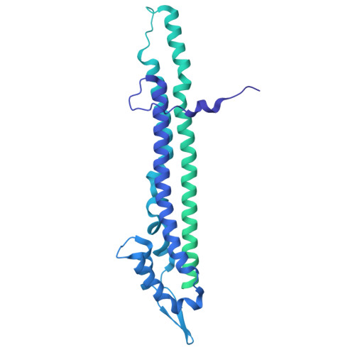
    8VVI, 8VVN

  • PubMed Abstract: 

    Bacteria have evolved an extraordinary diversity of defense systems against bacteriophage (phage) predation. However, the molecular mechanisms underlying these anti-phage systems often remain elusive. Here, we provide mechanistic and structural insights into Zorya phage defense systems. Using cryo-EM structural analyses, we show that the Zorya type I and II core components, ZorA and ZorB, assemble in a 5:2 complex that is similar to inner-membrane ion-driven, rotary motors that power flagellar rotation, type 9 secretion, gliding and the Ton nutrient uptake systems. The ZorAB complex has an elongated cytoplasmic tail assembled by bundling the C-termini of the five ZorA subunits. Mutagenesis demonstrates that peptidoglycan binding by the periplasmic domains of ZorB, the structured cytoplasmic tail of ZorA, and ion flow through the motor is important for function in both type I and II systems. Furthermore, we identify ZorE as the effector module of the Zorya II system, possessing nickase activity. Our work reveals the molecular basis of the activity of Zorya systems and highlights the ZorE nickase as crucial for population-wide immunity in the type II system.


  • Organizational Affiliation
    • Department of Microbial Sciences, Faculty of Health and Medical Sciences, University of Glasgow, Guildford, UK. giusy.mariano@glasgow.ac.uk.

Macromolecules
Find similar proteins by:  (by identity cutoff)  |  3D Structure
Entity ID: 1
MoleculeChains Sequence LengthOrganismDetailsImage
Chemotaxis protein MotB-related proteinA [auth B],
B [auth A]
282Shewanella sp. ANA-3Mutation(s): 0 
Gene Names: Shewana3_3786
Membrane Entity: Yes 
UniProt
Find proteins for A0L1T5 (Shewanella sp. (strain ANA-3))
Explore A0L1T5 
Go to UniProtKB:  A0L1T5
Entity Groups  
Sequence Clusters30% Identity50% Identity70% Identity90% Identity95% Identity100% Identity
UniProt GroupA0L1T5
Sequence Annotations
Expand
  • Reference Sequence
Find similar proteins by:  (by identity cutoff)  |  3D Structure
Entity ID: 2
MoleculeChains Sequence LengthOrganismDetailsImage
MotA/TolQ/ExbB proton channel domain-containing protein
C, D, E, F, G
696Shewanella sp. ANA-3Mutation(s): 0 
Gene Names: Shewana3_3785
Membrane Entity: Yes 
UniProt
Find proteins for A0L1T4 (Shewanella sp. (strain ANA-3))
Explore A0L1T4 
Go to UniProtKB:  A0L1T4
Entity Groups  
Sequence Clusters30% Identity50% Identity70% Identity90% Identity95% Identity100% Identity
UniProt GroupA0L1T4
Sequence Annotations
Expand
  • Reference Sequence
Experimental Data & Validation

Experimental Data

  • Method: ELECTRON MICROSCOPY
  • Resolution: 2.20 Å
  • Aggregation State: PARTICLE 
  • Reconstruction Method: SINGLE PARTICLE 
EM Software:
TaskSoftware PackageVersion
MODEL REFINEMENTPHENIX

Structure Validation

View Full Validation Report


Entry History & Funding Information

Deposition Data


Funding OrganizationLocationGrant Number
National Institutes of Health/National Cancer Institute (NIH/NCI)United StatesCCR Core Funding for Lea Group

Revision History  (Full details and data files)

  • Version 1.0: 2025-02-05
    Type: Initial release
  • Version 1.1: 2025-03-19
    Changes: Data collection, Database references
  • Version 1.2: 2025-06-04
    Changes: Data collection