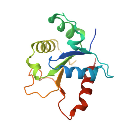
8W8M | pdb_00008w8m

Cryo-EM structure of helical filament of MyD88 TIR


Experimental Data Snapshot

  • Method: ELECTRON MICROSCOPY
  • Resolution: 3.28 Å
  • Aggregation State: HELICAL ARRAY 
  • Reconstruction Method: SINGLE PARTICLE 

wwPDB Validation   3D Report Full Report


This is version 1.1 of the entry. See complete history


Literature

From Monomers to Oligomers: Structural Mechanism of Receptor-Triggered MyD88 Assembly in Innate Immune Signaling

Kasai, K.Imamura, K.Uno, M.Sekiyama, N.Miyata, T.Makino, F.Yamada, R.Takahashi, Y.Kodera, N.Namba, K.Ohnishi, H.Narita, A.Konno, H.Tochio, H.

(2024) bioRxiv 


Macromolecules
Find similar proteins by:  (by identity cutoff)  |  3D Structure
Entity ID: 1
MoleculeChains Sequence LengthOrganismDetailsImage
Myeloid differentiation primary response protein MyD88144Homo sapiensMutation(s): 0 
Gene Names: MYD88
UniProt & NIH Common Fund Data Resources
Find proteins for Q99836 (Homo sapiens)
Explore Q99836 
Go to UniProtKB:  Q99836
PHAROS:  Q99836
GTEx:  ENSG00000172936 
Entity Groups  
Sequence Clusters30% Identity50% Identity70% Identity90% Identity95% Identity100% Identity
UniProt GroupQ99836
Sequence Annotations
Expand
  • Reference Sequence
Experimental Data & Validation

Experimental Data

  • Method: ELECTRON MICROSCOPY
  • Resolution: 3.28 Å
  • Aggregation State: HELICAL ARRAY 
  • Reconstruction Method: SINGLE PARTICLE 

Structure Validation

View Full Validation Report


Entry History & Funding Information

Deposition Data


Funding OrganizationLocationGrant Number
Japan Science and TechnologyJapanJPMJCR1762
Japan Society for the Promotion of Science (JSPS)Japan16H04752

Revision History  (Full details and data files)

  • Version 1.0: 2024-09-04
    Type: Initial release
  • Version 1.1: 2025-03-26
    Changes: Data collection, Database references, Structure summary