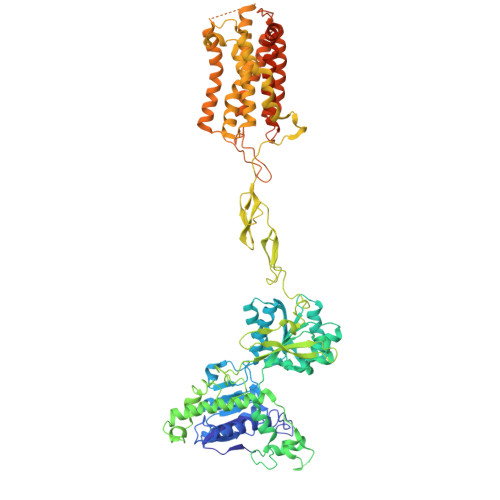
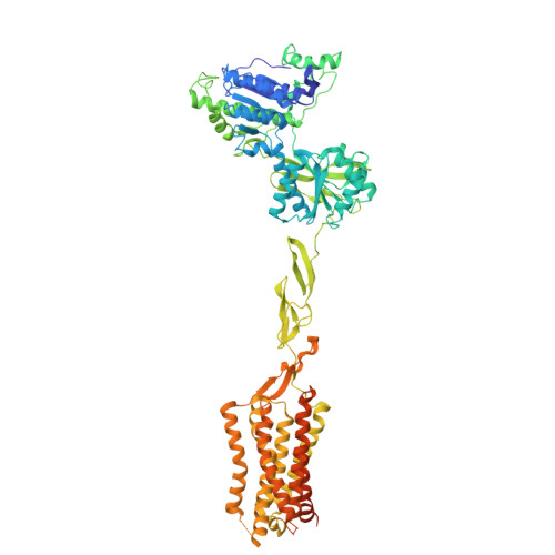
8WGD | pdb_00008wgd

mGlu2-4 inactive heterodimer


Experimental Data Snapshot

  • Method: ELECTRON MICROSCOPY
  • Resolution: 4.45 Å
  • Aggregation State: PARTICLE 
  • Reconstruction Method: SINGLE PARTICLE 

wwPDB Validation   3D Report Full Report


This is version 1.1 of the entry. See complete history


Literature

Structural basis of orientated asymmetry in a mGlu heterodimer.

Huang, W.Jin, N.Guo, J.Shen, C.Xu, C.Xi, K.Bonhomme, L.Quast, R.B.Shen, D.D.Qin, J.Liu, Y.R.Song, Y.Gao, Y.Margeat, E.Rondard, P.Pin, J.P.Zhang, Y.Liu, J.

(2024) Nat Commun 15: 10345-10345

  • DOI: https://doi.org/10.1038/s41467-024-54744-7
  • Primary Citation of Related Structures:  
    8WG9, 8WGB, 8WGC, 8WGD

  • PubMed Abstract: 

    The structural basis for the allosteric interactions within G protein-coupled receptors (GPCRs) heterodimers remains largely unknown. The metabotropic glutamate (mGlu) receptors are complex dimeric GPCRs important for the fine tuning of many synapses. Heterodimeric mGlu receptors with specific allosteric properties have been identified in the brain. Here we report four cryo-electron microscopy structures of mGlu2-4 heterodimer in different states: an inactive state bound to antagonists, two intermediate states bound to either mGlu2 or mGlu4 agonist only and an active state bound to both glutamate and a mGlu4 positive allosteric modulator (PAM) in complex with Gi protein. In addition to revealing a unique PAM binding pocket among mGlu receptors, our data bring important information for the asymmetric activation of mGlu heterodimers. First, we show that agonist binding to a single subunit in the extracellular domain is not sufficient to stabilize an active dimer conformation. Single-molecule FRET data show that the monoliganded mGlu2-4 can be found in both intermediate states and an active one. Second, we provide a detailed view of the asymmetric interface in seven-transmembrane (7TM) domains and identified key residues within the mGlu2 7TM that limits its activation leaving mGlu4 as the only subunit activating G proteins.


  • Organizational Affiliation
    • Key Laboratory of Molecular Biophysics of MOE, College of Life Science and Technology, Huazhong University of Science and Technology (HUST), Wuhan, China.

Macromolecules
Find similar proteins by:  (by identity cutoff)  |  3D Structure
Entity ID: 1
MoleculeChains Sequence LengthOrganismDetailsImage
Metabotropic glutamate receptor 2A [auth C]854Homo sapiensMutation(s): 0 
Gene Names: GRM2
Membrane Entity: Yes 
UniProt & NIH Common Fund Data Resources
Find proteins for Q14416 (Homo sapiens)
Explore Q14416 
Go to UniProtKB:  Q14416
PHAROS:  Q14416
GTEx:  ENSG00000164082 
Entity Groups  
Sequence Clusters30% Identity50% Identity70% Identity90% Identity95% Identity100% Identity
UniProt GroupQ14416
Sequence Annotations
Expand
  • Reference Sequence
Find similar proteins by:  (by identity cutoff)  |  3D Structure
Entity ID: 2
MoleculeChains Sequence LengthOrganismDetailsImage
Metabotropic glutamate receptor 4880Homo sapiensMutation(s): 0 
Gene Names: GRM4
Membrane Entity: Yes 
UniProt & NIH Common Fund Data Resources
Find proteins for Q14833 (Homo sapiens)
Explore Q14833 
Go to UniProtKB:  Q14833
PHAROS:  Q14833
GTEx:  ENSG00000124493 
Entity Groups  
Sequence Clusters30% Identity50% Identity70% Identity90% Identity95% Identity100% Identity
UniProt GroupQ14833
Sequence Annotations
Expand
  • Reference Sequence
Small Molecules
Ligands 2 Unique
IDChains Name / Formula / InChI Key2D Diagram3D Interactions
WAG (Subject of Investigation/LOI)
Query on WAG

Download Ideal Coordinates CCD File 
C
(1S,2R)-2-[(2S)-2-azanyl-1-oxidanyl-1-oxidanylidene-3-(9H-xanthen-9-yl)propan-2-yl]cyclopropane-1-carboxylic acid
C20 H19 N O5
VLZBRVJVCCNPRJ-AGKSTNPPSA-N
WA6 (Subject of Investigation/LOI)
Query on WA6

Download Ideal Coordinates CCD File 
D [auth B](2S)-2-azanyl-2-cyclopropyl-2-(4-phosphonophenyl)ethanoic acid
C11 H14 N O5 P
IGODGTDUQSMDQU-NSHDSACASA-N
Experimental Data & Validation

Experimental Data

  • Method: ELECTRON MICROSCOPY
  • Resolution: 4.45 Å
  • Aggregation State: PARTICLE 
  • Reconstruction Method: SINGLE PARTICLE 

Structure Validation

View Full Validation Report


Entry History & Funding Information

Deposition Data


Funding OrganizationLocationGrant Number
National Natural Science Foundation of China (NSFC)China--

Revision History  (Full details and data files)

  • Version 1.0: 2024-11-27
    Type: Initial release
  • Version 1.1: 2025-05-28
    Changes: Data collection, Database references