8X3M | pdb_00008x3m

Crystal structure of p38alpha with an allosteric inhibitor 2


Experimental Data Snapshot

  • Method: X-RAY DIFFRACTION
  • Resolution: 1.85 Å
  • R-Value Free: 
    0.226 (Depositor), 0.225 (DCC) 
  • R-Value Work: 
    0.181 (Depositor), 0.180 (DCC) 
  • R-Value Observed: 
    0.183 (Depositor) 

Starting Model: experimental
View more details

wwPDB Validation   3D Report Full Report


Ligand Structure Quality Assessment 


This is version 1.1 of the entry. See complete history


Literature

Distinct binding modes of a benzothiazole derivative confer structural bases for increasing ERK2 or p38 alpha MAPK selectivity.

Hasegawa, S.Yoshida, M.Nagao, H.Sugiyama, H.Sawa, M.Kinoshita, T.

(2024) Biochem Biophys Res Commun 704: 149707-149707

  • DOI: https://doi.org/10.1016/j.bbrc.2024.149707
  • Primary Citation of Related Structures:  
    8X3M

  • PubMed Abstract: 

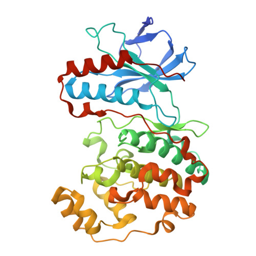
    Mitogen-activated protein kinases (MAPKs), including extracellular signal-regulated kinase 2 (ERK2) and p38α MAP kinase (p38α MAPK), regulate various cellular responses. ERK2 is a drug target for treating many diseases, such as cancer, whereas p38α has attracted much attention as a promising drug target for treating inflammatory disorders. ERK2 is a critical off-target for p38α MAPK and vice versa. In this study, an allosteric ERK2 inhibitor with a benzothiazole moiety (compound 1) displayed comparable inhibitory activity against p38α MAPK. Crystal structures of these MAPKs showed that compound 1 bound to the allosteric site of ERK2 and p38α MAPK in distinct manners. Compound 1 formed a covalent bond with Cys162 of p38α MAPK, whereas this covalent bond was absent in the ERK2 complex even though the corresponding cysteine is conserved in ERK2. Structural dissection combined with computational simulations indicated that an amino acid difference in the allosteric site is responsible for the distinct binding modes of compound 1 with ERK2 and p38α MAPK. These structural insights underline the feasibility of developing highly selective and potent ERK2 and p38α MAPK inhibitors.


  • Organizational Affiliation

    Graduate School of Science, Osaka Metropolitan University, Osaka, 599-8570, Japan.


Macromolecules
Find similar proteins by:  (by identity cutoff)  |  3D Structure
Entity ID: 1
MoleculeChains Sequence LengthOrganismDetailsImage
Mitogen-activated protein kinase 14365Homo sapiensMutation(s): 0 
Gene Names: MAPK14CSBPCSBP1CSBP2CSPB1MXI2SAPK2A
EC: 2.7.11.24
UniProt & NIH Common Fund Data Resources
Find proteins for Q16539 (Homo sapiens)
Explore Q16539 
Go to UniProtKB:  Q16539
PHAROS:  Q16539
GTEx:  ENSG00000112062 
Entity Groups  
Sequence Clusters30% Identity50% Identity70% Identity90% Identity95% Identity100% Identity
UniProt GroupQ16539
Sequence Annotations
Expand
  • Reference Sequence
Small Molecules
Ligands 1 Unique
IDChains Name / Formula / InChI Key2D Diagram3D Interactions
B8Z
Query on B8Z

Download Ideal Coordinates CCD File 
B [auth A]~{N}-(5,6-dimethoxy-1,3-benzothiazol-2-yl)-2-[(4-fluoranylphenoxy)methyl]-1,3-thiazole-4-carboxamide
C20 H16 F N3 O4 S2
PCKXVDUIUFNAJD-UHFFFAOYSA-N
Experimental Data & Validation

Experimental Data

  • Method: X-RAY DIFFRACTION
  • Resolution: 1.85 Å
  • R-Value Free:  0.226 (Depositor), 0.225 (DCC) 
  • R-Value Work:  0.181 (Depositor), 0.180 (DCC) 
  • R-Value Observed: 0.183 (Depositor) 
Space Group: P 21 21 21
Unit Cell:
Length ( Å )Angle ( ˚ )
a = 45.366α = 90
b = 85.5β = 90
c = 125.669γ = 90
Software Package:
Software NamePurpose
PHENIXrefinement
PHENIXrefinement
XDSdata reduction
XDSdata scaling
PHASERphasing

Structure Validation

View Full Validation Report


Ligand Structure Quality Assessment 


Entry History & Funding Information

Deposition Data


Funding OrganizationLocationGrant Number
Not funded--

Revision History  (Full details and data files)

  • Version 1.0: 2024-11-20
    Type: Initial release
  • Version 1.1: 2025-03-05
    Changes: Database references