8YVB | pdb_00008yvb

Crystal structure of Caenorhabditis elegans ZIM-1 ZF1-2-CTD domain in complex with Chromosome II/III pairing center


Experimental Data Snapshot

  • Method: X-RAY DIFFRACTION
  • Resolution: 1.90 Å
  • R-Value Free: 
    0.239 (Depositor), 0.190 (DCC) 
  • R-Value Work: 
    0.190 (Depositor), 0.192 (DCC) 
  • R-Value Observed: 
    0.192 (Depositor) 

wwPDB Validation   3D Report Full Report


This is version 1.0 of the entry. See complete history


Literature

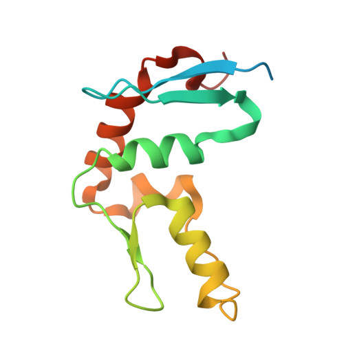
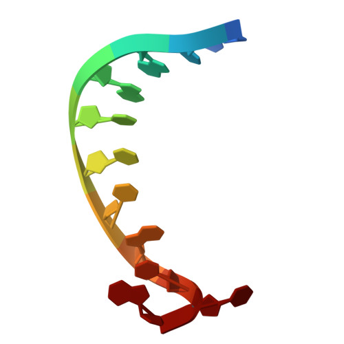
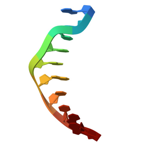
Structural basis for C. elegans pairing center DNA binding specificity by the ZIM/HIM-8 family proteins.

Li, M.Zhu, C.Xu, Z.Xu, M.Kuang, Y.Hou, X.Huang, X.Lv, M.Liu, Y.Zhang, Y.Xu, Z.Han, X.Wang, S.Shi, Y.Guang, S.Li, F.

(2024) Nat Commun 15: 10355-10355

  • DOI: https://doi.org/10.1038/s41467-024-54548-9
  • Primary Citation of Related Structures:  
    8YV9, 8YVA, 8YVB

  • PubMed Abstract: 

    Pairing center (PC) on each chromosome of Caenorhabditis elegans is crucial for homolog pairing and initiating synapsis. Within each PC, clusters of 11/12 bp DNA motif recruit one of four paralogous meiosis-specific proteins: ZIM-1, ZIM-2, ZIM-3, or HIM-8. However, the mechanistic basis underlying the specificity of ZIM/HIM-8-PC DNA interaction remains elusive. Here, we describe crystal structures of HIM-8, ZIM-1 and ZIM-2 DNA binding domains (ZF1, ZF2 and CTD) in complex with their cognate PC DNA motifs, respectively. These structures demonstrated the ZF1-2-CTD folds as an integrated structural unit crucial for its DNA binding specificity. Base-specific DNA-contacting residues are exclusively distributed on ZF1-2 and highly conserved. Furthermore, the CTD potentially contributes to the conformational diversity of ZF1-2, imparting binding specificity to distinct PC DNA motifs. These findings shed light on the mechanism governing PC DNA motif recognition by ZIM/HIM-8 proteins, suggesting a co-evolution relationship between PC DNA motifs and ZF1-2-CTD in shaping the specific recognition.


  • Organizational Affiliation
    • MOE Key Laboratory for Cellular Dynamics, Hefei National Laboratory for Physical Sciences at the Microscale, The First Affiliated Hospital of USTC, Biomedical Sciences and Health Laboratory of Anhui Province, Division of Life Sciences and Medicine, University of Science and Technology of China, Hefei, China.

Macromolecules

Find similar proteins by:  (by identity cutoff)  |  3D Structure
Entity ID: 1
MoleculeChains Sequence LengthOrganismDetailsImage
C2H2-type domain-containing protein128Caenorhabditis elegansMutation(s): 0 
Gene Names: zim-1CELE_T07G12.6T07G12.6
UniProt
Find proteins for B2MZC6 (Caenorhabditis elegans)
Explore B2MZC6 
Go to UniProtKB:  B2MZC6
Entity Groups  
Sequence Clusters30% Identity50% Identity70% Identity90% Identity95% Identity100% Identity
UniProt GroupB2MZC6
Sequence Annotations
Expand
  • Reference Sequence

Find similar nucleic acids by:  Sequence   |   3D Structure  

Entity ID: 2
MoleculeChains LengthOrganismImage
DNA (5'-D(*TP*TP*GP*GP*TP*CP*TP*GP*CP*TP*A)-3')11Caenorhabditis elegans
Sequence Annotations
Expand
  • Reference Sequence

Find similar nucleic acids by:  Sequence   |   3D Structure  

Entity ID: 3
MoleculeChains LengthOrganismImage
DNA (5'-D(P*AP*GP*CP*AP*GP*AP*CP*CP*AP*A)-3')10Caenorhabditis elegans
Sequence Annotations
Expand
  • Reference Sequence
Experimental Data & Validation

Experimental Data

  • Method: X-RAY DIFFRACTION
  • Resolution: 1.90 Å
  • R-Value Free:  0.239 (Depositor), 0.190 (DCC) 
  • R-Value Work:  0.190 (Depositor), 0.192 (DCC) 
  • R-Value Observed: 0.192 (Depositor) 
Space Group: C 2 2 21
Unit Cell:
Length ( Å )Angle ( ˚ )
a = 55.767α = 90
b = 62.464β = 90
c = 101.564γ = 90
Software Package:
Software NamePurpose
PHENIXrefinement
HKL-2000data scaling
HKL-2000data reduction
PHENIXphasing

Structure Validation

View Full Validation Report


Entry History & Funding Information

Deposition Data


Funding OrganizationLocationGrant Number
National Natural Science Foundation of China (NSFC)China32371282
National Natural Science Foundation of China (NSFC)China32171222

Revision History  (Full details and data files)

  • Version 1.0: 2024-12-11
    Type: Initial release