


A sensitive sensor for discovering honokiol as a potent STING activator that enhances antitumor immunity
Sun, P.K., Li, X.J.To be published.
Experimental Data Snapshot
Entity ID: 1 | |||||
|---|---|---|---|---|---|
| Molecule | Chains | Sequence Length | Organism | Details | Image |
| Stimulator of interferon genes protein | 192 | Homo sapiens | Mutation(s): 0 Gene Names: STING1, ERIS, MITA, STING, TMEM173 | 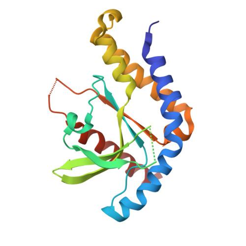 | |
UniProt & NIH Common Fund Data Resources | |||||
Find proteins for Q86WV6 (Homo sapiens) Explore Q86WV6 Go to UniProtKB: Q86WV6 | |||||
PHAROS: Q86WV6 GTEx: ENSG00000184584 | |||||
Entity Groups | |||||
| Sequence Clusters | 30% Identity50% Identity70% Identity90% Identity95% Identity100% Identity | ||||
| UniProt Group | Q86WV6 | ||||
Sequence AnnotationsExpand | |||||
| |||||
| Ligands 3 Unique | |||||
|---|---|---|---|---|---|
| ID | Chains | Name / Formula / InChI Key | 2D Diagram | 3D Interactions | |
| Y4T (Subject of Investigation/LOI) Query on Y4T | D [auth A] | (1P)-3',5-di(prop-2-en-1-yl)[1,1'-biphenyl]-2,4'-diol C18 H18 O2 FVYXIJYOAGAUQK-UHFFFAOYSA-N |  | ||
| CA Query on CA | C [auth A] | CALCIUM ION Ca BHPQYMZQTOCNFJ-UHFFFAOYSA-N |  | ||
| CL Query on CL | B [auth A] | CHLORIDE ION Cl VEXZGXHMUGYJMC-UHFFFAOYSA-M |  | ||
| Length ( Å ) | Angle ( ˚ ) |
|---|---|
| a = 80.714 | α = 90 |
| b = 89.477 | β = 90 |
| c = 72.955 | γ = 90 |
| Software Name | Purpose |
|---|---|
| PHENIX | refinement |
| XDS | data scaling |
| XDS | data reduction |
| PHASER | phasing |
| Funding Organization | Location | Grant Number |
|---|---|---|
| National Natural Science Foundation of China (NSFC) | China | 92157104 |


