8Z3R | pdb_00008z3r

The structure of type III CRISPR-associated deaminase in complex cA4


Experimental Data Snapshot

  • Method: ELECTRON MICROSCOPY
  • Resolution: 2.28 Å
  • Aggregation State: PARTICLE 
  • Reconstruction Method: SINGLE PARTICLE 

wwPDB Validation   3D Report Full Report


This is version 1.2 of the entry. See complete history


Literature

Antiviral signaling of a type III CRISPR-associated deaminase.

Li, Y.Li, Z.Yan, P.Hua, C.Kong, J.Wu, W.Cui, Y.Duan, Y.Li, S.Li, G.Ji, S.Chen, Y.Zhao, Y.Yang, P.Hu, C.Lu, M.Chen, M.Xiao, Y.

(2025) Science 387: eadr0393-eadr0393

  • DOI: https://doi.org/10.1126/science.adr0393
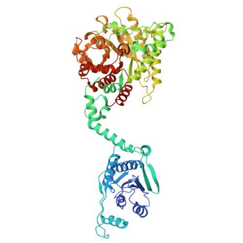
  • Primary Citation of Related Structures:  
    8Z3K, 8Z3P, 8Z3R, 8Z40

  • PubMed Abstract: 

    Prokaryotes have evolved diverse defense strategies against viral infection, such as foreign nucleic acid degradation by CRISPR-Cas systems and DNA/RNA synthesis inhibition via nucleotide pool depletion. Here, we report an antiviral mechanism of type III CRISPR-Cas-regulated ATP depletion, where ATP is converted into ITP by CRISPR-Cas-associated adenosine deaminase (CAAD) upon activation by either cA 4 or cA 6 , followed by hydrolysis into IMP by Nudix hydrolase, ultimately resulting in cell growth arrest. The cryo-electron microscopy structures of CAAD in its apo and activated forms, together with biochemical evidence, revealed how cA 4 /cA 6 binds to the CARF domain and abrogates CAAD autoinhibition, inducing substantial conformational changes that reshape the structure of CAAD and induce its deaminase activity. Our results reveal the mechanism of a CRISPR-Cas-regulated ATP depletion antiviral strategy.


  • Organizational Affiliation
    • Department of Pharmacology, School of Pharmacy, China Pharmaceutical University, Nanjing, China.

Macromolecules
Find similar proteins by:  (by identity cutoff)  |  3D Structure
Entity ID: 1
MoleculeChains Sequence LengthOrganismDetailsImage
Adenosine deaminase domain-containing protein
A, B, C, D, E
A, B, C, D, E, F
635Limisphaera ngatamarikiensisMutation(s): 0 
Gene Names: G4L39_03315
EC: 3.5.4.4
UniProt
Find proteins for A0A6M1RED6 (Limisphaera ngatamarikiensis)
Explore A0A6M1RED6 
Go to UniProtKB:  A0A6M1RED6
Entity Groups  
Sequence Clusters30% Identity50% Identity70% Identity90% Identity95% Identity100% Identity
UniProt GroupA0A6M1RED6
Sequence Annotations
Expand
  • Reference Sequence
Small Molecules
Ligands 2 Unique
IDChains Name / Formula / InChI Key2D Diagram3D Interactions
LQJ
Query on LQJ

Download Ideal Coordinates CCD File 
H [auth A]
J [auth B]
L [auth C]
N [auth D]
Q [auth F]
H [auth A],
J [auth B],
L [auth C],
N [auth D],
Q [auth F],
R [auth F]
3'-O-[(R)-{[(2S,3aS,4S,6S,6aS)-6-(6-amino-9H-purin-9-yl)-2-hydroxy-2-oxotetrahydro-2H-2lambda~5~-furo[3,4-d][1,3,2]dioxaphosphol-4-yl]methoxy}(hydroxy)phosphoryl]adenosine
C20 H24 N10 O12 P2
ZKDCIJKZCQKMKS-RJLGOGMOSA-N
ZN (Subject of Investigation/LOI)
Query on ZN

Download Ideal Coordinates CCD File 
G [auth A]
I [auth B]
K [auth C]
M [auth D]
O [auth E]
G [auth A],
I [auth B],
K [auth C],
M [auth D],
O [auth E],
P [auth F]
ZINC ION
Zn
PTFCDOFLOPIGGS-UHFFFAOYSA-N
Experimental Data & Validation

Experimental Data

  • Method: ELECTRON MICROSCOPY
  • Resolution: 2.28 Å
  • Aggregation State: PARTICLE 
  • Reconstruction Method: SINGLE PARTICLE 
EM Software:
TaskSoftware PackageVersion
MODEL REFINEMENTPHENIX

Structure Validation

View Full Validation Report


Entry History & Funding Information

Deposition Data


Funding OrganizationLocationGrant Number
National Natural Science Foundation of China (NSFC)China--

Revision History  (Full details and data files)

  • Version 1.0: 2024-12-25
    Type: Initial release
  • Version 1.1: 2025-03-05
    Changes: Data collection, Database references
  • Version 1.2: 2025-07-23
    Changes: Data collection, Database references