8BPI | pdb_00008bpi

Human Gamma-D crystallin R36S mutant after UV illumination


Experimental Data Snapshot

  • Method: X-RAY DIFFRACTION
  • Resolution: 2.00 Å
  • R-Value Free: 
    0.241 (Depositor), 0.250 (DCC) 
  • R-Value Work: 
    0.197 (Depositor), 0.200 (DCC) 

wwPDB Validation   3D Report Full Report


This is version 1.2 of the entry. See complete history


Literature

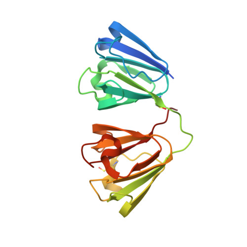
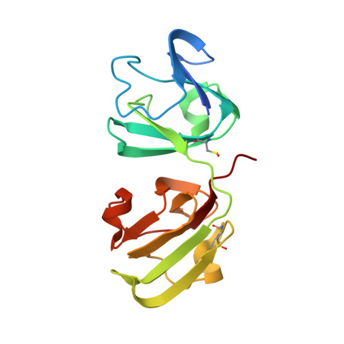
An ultraviolet-driven rescue pathway for oxidative stress to eye lens protein human gamma-D crystallin.

Hill, J.A.Nyathi, Y.Horrell, S.von Stetten, D.Axford, D.Owen, R.L.Beddard, G.S.Pearson, A.R.Ginn, H.M.Yorke, B.A.

(2024) Commun Chem 7: 81-81

  • DOI: https://doi.org/10.1038/s42004-024-01163-w
  • Primary Citation of Related Structures:  
    8BD0, 8BPI, 8Q3L

  • PubMed Abstract: 

    Human gamma-D crystallin (HGD) is a major constituent of the eye lens. Aggregation of HGD contributes to cataract formation, the leading cause of blindness worldwide. It is unique in its longevity, maintaining its folded and soluble state for 50-60 years. One outstanding question is the structural basis of this longevity despite oxidative aging and environmental stressors including ultraviolet radiation (UV). Here we present crystallographic structures evidencing a UV-induced crystallin redox switch mechanism. The room-temperature serial synchrotron crystallographic (SSX) structure of freshly prepared crystallin mutant (R36S) shows no post-translational modifications. After aging for nine months in the absence of light, a thiol-adduct (dithiothreitol) modifying surface cysteines is observed by low-dose SSX. This is shown to be UV-labile in an acutely light-exposed structure. This suggests a mechanism by which a major source of crystallin damage, UV, may also act as a rescuing factor in a finely balanced redox system.


  • Organizational Affiliation
    • School of Chemistry and Biosciences, University of Bradford, Richmond Road, Bradford, BD7 1DP, United Kingdom.

Macromolecules
Find similar proteins by:  (by identity cutoff)  |  3D Structure
Entity ID: 1
MoleculeChains Sequence LengthOrganismDetailsImage
Gamma-crystallin DA [auth XXX]182Homo sapiensMutation(s): 1 
Gene Names: CRYGDCRYG4
UniProt & NIH Common Fund Data Resources
Find proteins for P07320 (Homo sapiens)
Explore P07320 
Go to UniProtKB:  P07320
PHAROS:  P07320
GTEx:  ENSG00000118231 
Entity Groups  
Sequence Clusters30% Identity50% Identity70% Identity90% Identity95% Identity100% Identity
UniProt GroupP07320
Sequence Annotations
Expand
  • Reference Sequence
Find similar proteins by:  (by identity cutoff)  |  3D Structure
Entity ID: 2
MoleculeChains Sequence LengthOrganismDetailsImage
Gamma-crystallin DB [auth AAA]182Homo sapiensMutation(s): 1 
Gene Names: CRYGDCRYG4
UniProt & NIH Common Fund Data Resources
Find proteins for P07320 (Homo sapiens)
Explore P07320 
Go to UniProtKB:  P07320
PHAROS:  P07320
GTEx:  ENSG00000118231 
Entity Groups  
Sequence Clusters30% Identity50% Identity70% Identity90% Identity95% Identity100% Identity
UniProt GroupP07320
Sequence Annotations
Expand
  • Reference Sequence
Small Molecules
Modified Residues  1 Unique
IDChains TypeFormula2D DiagramParent
CSO
Query on CSO
A [auth XXX]L-PEPTIDE LINKINGC3 H7 N O3 SCYS
Experimental Data & Validation

Experimental Data

  • Method: X-RAY DIFFRACTION
  • Resolution: 2.00 Å
  • R-Value Free:  0.241 (Depositor), 0.250 (DCC) 
  • R-Value Work:  0.197 (Depositor), 0.200 (DCC) 
Space Group: P 21 21 21
Unit Cell:
Length ( Å )Angle ( ˚ )
a = 53.9α = 90
b = 83.7β = 90
c = 101γ = 90
Software Package:
Software NamePurpose
REFMACrefinement
CrystFELdata reduction
CrystFELdata scaling
MOLREPphasing

Structure Validation

View Full Validation Report


Entry History & Funding Information

Deposition Data


Funding OrganizationLocationGrant Number
Wellcome TrustUnited KingdomSBF0061044

Revision History  (Full details and data files)

  • Version 1.0: 2023-11-29
    Type: Initial release
  • Version 1.1: 2024-08-14
    Changes: Database references
  • Version 1.2: 2024-11-20
    Changes: Structure summary