9BSV | pdb_00009bsv

EBOV GP/Nanosota-EB2 complex


Experimental Data Snapshot

  • Method: ELECTRON MICROSCOPY
  • Resolution: 3.10 Å
  • Aggregation State: PARTICLE 
  • Reconstruction Method: SINGLE PARTICLE 

Starting Model: experimental
View more details

wwPDB Validation   3D Report Full Report


This is version 1.1 of the entry. See complete history


Literature

Discovery of Nanosota-EB1 and -EB2 as Novel Nanobody Inhibitors Against Ebola Virus Infection.

Bu, F.Ye, G.Morsheimer, K.Mendoza, A.Turner-Hubbard, H.Herbst, M.Spiller, B.Wadzinski, B.E.Eaton, B.Anantpadma, M.Yang, G.Liu, B.Davey, R.Li, F.

(2024) PLoS Pathog 20: e1012817-e1012817

  • DOI: https://doi.org/10.1371/journal.ppat.1012817
  • Primary Citation of Related Structures:  
    9BSU, 9BSV

  • PubMed Abstract: 

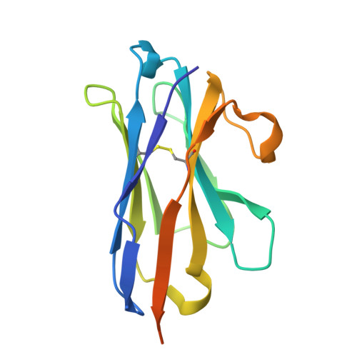
    The Ebola filovirus (EBOV) poses a serious threat to global health and national security. Nanobodies, a type of single-domain antibody, have demonstrated promising therapeutic potential. We identified two anti-EBOV nanobodies, Nanosota-EB1 and Nanosota-EB2, which specifically target the EBOV glycoprotein (GP). Cryo-EM and biochemical data revealed that Nanosota-EB1 binds to the glycan cap of GP1, preventing its protease cleavage, while Nanosota-EB2 binds to critical membrane-fusion elements in GP2, stabilizing it in the pre-fusion state. Nanosota-EB2 is a potent neutralizer of EBOV infection in vitro and offers excellent protection in a mouse model of EBOV challenge, while Nanosota-EB1 provides moderate neutralization and protection. Nanosota-EB1 and Nanosota-EB2 are the first nanobodies shown to inhibit authentic EBOV. Combined with our newly developed structure-guided in vitro evolution approach, they lay the foundation for nanobody-based therapies against EBOV and other viruses within the ebolavirus genus.


  • Organizational Affiliation
    • Department of Pharmacology, University of Minnesota Medical School, Minneapolis, Minnesota, United States of America.

Macromolecules
Find similar proteins by:  (by identity cutoff)  |  3D Structure
Entity ID: 1
MoleculeChains Sequence LengthOrganismDetailsImage
Nanosota-EB2A [auth D],
E,
F
144Vicugna pacosMutation(s): 0 
Entity Groups  
Sequence Clusters30% Identity50% Identity70% Identity90% Identity95% Identity100% Identity
Sequence Annotations
Expand
  • Reference Sequence
Find similar proteins by:  (by identity cutoff)  |  3D Structure
Entity ID: 2
MoleculeChains Sequence LengthOrganismDetailsImage
Envelope glycoproteinB [auth A],
C [auth B],
D [auth C]
706Ebola virusMutation(s): 0 
Gene Names: GP
UniProt
Find proteins for Q05320 (Zaire ebolavirus (strain Mayinga-76))
Explore Q05320 
Go to UniProtKB:  Q05320
Entity Groups  
Sequence Clusters30% Identity50% Identity70% Identity90% Identity95% Identity100% Identity
UniProt GroupQ05320
Glycosylation
Glycosylation Sites: 1
Sequence Annotations
Expand
  • Reference Sequence
Oligosaccharides

Help

Entity ID: 3
MoleculeChains Length2D Diagram Glycosylation3D Interactions
alpha-D-mannopyranose-(1-4)-[alpha-D-mannopyranose-(1-6)]alpha-D-mannopyranose-(1-6)-[alpha-D-mannopyranose-(1-3)]alpha-D-mannopyranose-(1-4)-2-acetamido-2-deoxy-beta-D-glucopyranose-(1-4)-2-acetamido-2-deoxy-beta-D-glucopyranose
G
7N-Glycosylation
Entity ID: 4
MoleculeChains Length2D Diagram Glycosylation3D Interactions
alpha-D-mannopyranose-(1-2)-[beta-D-mannopyranose-(1-6)]alpha-D-mannopyranose-(1-6)-[alpha-D-mannopyranose-(1-3)]beta-D-mannopyranose-(1-4)-2-acetamido-2-deoxy-beta-D-glucopyranose-(1-4)-2-acetamido-2-deoxy-beta-D-glucopyranose
H
7N-Glycosylation
Entity ID: 5
MoleculeChains Length2D Diagram Glycosylation3D Interactions
alpha-D-mannopyranose-(1-3)-alpha-D-mannopyranose-(1-6)-[alpha-D-mannopyranose-(1-3)]beta-D-mannopyranose-(1-4)-2-acetamido-2-deoxy-beta-D-glucopyranose-(1-4)-2-acetamido-2-deoxy-beta-D-glucopyranose
I
6N-Glycosylation
Glycosylation Resources
GlyTouCan:  G09724ZC
GlyCosmos:  G09724ZC
GlyGen:  G09724ZC
Small Molecules
Ligands 1 Unique
IDChains Name / Formula / InChI Key2D Diagram3D Interactions
MAN
Query on MAN

Download Ideal Coordinates CCD File 
J [auth E]alpha-D-mannopyranose
C6 H12 O6
WQZGKKKJIJFFOK-PQMKYFCFSA-N
Experimental Data & Validation

Experimental Data

  • Method: ELECTRON MICROSCOPY
  • Resolution: 3.10 Å
  • Aggregation State: PARTICLE 
  • Reconstruction Method: SINGLE PARTICLE 
EM Software:
TaskSoftware PackageVersion
MODEL REFINEMENTPHENIX1.21.1_5286:

Structure Validation

View Full Validation Report


Entry History & Funding Information

Deposition Data


Funding OrganizationLocationGrant Number
National Institutes of Health/National Institute Of Allergy and Infectious Diseases (NIH/NIAID)United StatesAI089728
National Institutes of Health/National Institute Of Allergy and Infectious Diseases (NIH/NIAID)United StatesAI110700
National Institutes of Health/National Institute Of Allergy and Infectious Diseases (NIH/NIAID)United StatesAI171954

Revision History  (Full details and data files)

  • Version 1.0: 2024-12-18
    Type: Initial release
  • Version 1.1: 2025-01-29
    Changes: Data collection, Database references