9C0X | pdb_00009c0x

Crystal structure of chimeric hemagglutinin cH11/1 in complex with broad protective antibody 31.b.09


Experimental Data Snapshot

  • Method: X-RAY DIFFRACTION
  • Resolution: 4.35 Å
  • R-Value Free: 
    0.382 (Depositor), 0.410 (DCC) 
  • R-Value Work: 
    0.335 (Depositor), 0.350 (DCC) 
  • R-Value Observed: 
    0.337 (Depositor) 

Starting Model: experimental
View more details

wwPDB Validation   3D Report Full Report


Ligand Structure Quality Assessment 


This is version 1.1 of the entry. See complete history


Literature

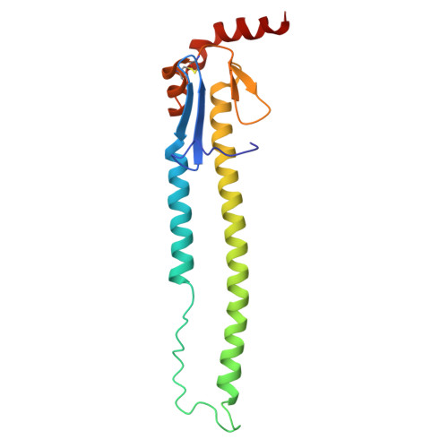
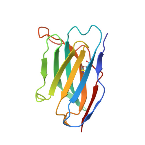
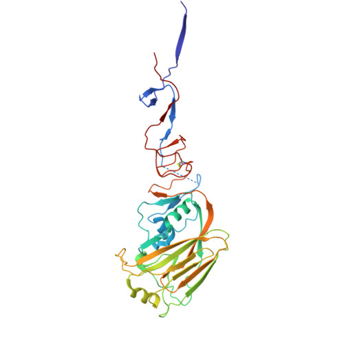
Structural characterization of influenza group 1 chimeric hemagglutinins as broad vaccine immunogens.

Nguyen, Y.T.K.Zhu, X.Han, J.Rodriguez, A.J.Sun, W.Yu, W.Palese, P.Krammer, F.Ward, A.B.Wilson, I.A.

(2025) Proc Natl Acad Sci U S A 122: e2416628122-e2416628122

  • DOI: https://doi.org/10.1073/pnas.2416628122
  • Primary Citation of Related Structures:  
    9C0U, 9C0V, 9C0X, 9C22

  • PubMed Abstract: 

    Chimeric hemagglutinins (cHA) appear to be promising for the design and development of universal influenza vaccines. Influenza A group 1 cHAs, cH5/1, cH8/1, and cH11/1, comprising an H1 stem attached to either an H5, H8, or H11 globular head, have been used sequentially as vaccine immunogens in human clinical trials and induced high levels of broadly protective antibodies. Using X-ray crystallography and negative-stain electron microscopy, we determined structures of cH5/1, cH8/1, and cH11/1 HAs in their apo (unliganded) and antibody Fab-bound states. Stem-reactive antibodies 3E1 and 31.b.09 recognize their cognate epitopes in cH5/1, cH8/1, and cH11/1 HAs. However, with cH5/1, the head domains are rotated by 35 to 45° around the threefold axis of the HA trimer compared to native HA with a more splayed-open conformation at the stem base. cH11/1 with 3E1 is structurally more native-like but resembles cH5/1 with 31.b.09, whereas cH8/1 with 31.b.09 exhibited a range of closed-to-open stem configurations with some separation of head and stem domains. Furthermore, all of these group 1 cHAs effectively bound a broad head trimer interface antibody and other broad stem antibodies. Thus, the cHAs exhibit structural plasticity without compromising the stem and head trimer interface epitopes for elicitation of influenza A group 1 cross-reactive antibodies.


  • Organizational Affiliation
    • Department of Integrative Structural and Computational Biology, The Scripps Research Institute, La Jolla, CA 92037.

Macromolecules
Find similar proteins by:  (by identity cutoff)  |  3D Structure
Entity ID: 1
MoleculeChains Sequence LengthOrganismDetailsImage
Hemagglutinin HA2 subunitA [auth B]166Influenza A virusMutation(s): 0 
Gene Names: HA
UniProt
Find proteins for A0A6J3XB93 (Influenza A virus)
Explore A0A6J3XB93 
Go to UniProtKB:  A0A6J3XB93
Entity Groups  
Sequence Clusters30% Identity50% Identity70% Identity90% Identity95% Identity100% Identity
UniProt GroupA0A6J3XB93
Sequence Annotations
Expand
  • Reference Sequence
Find similar proteins by:  (by identity cutoff)  |  3D Structure
Entity ID: 2
MoleculeChains Sequence LengthOrganismDetailsImage
Antibody 31.b.09 Fab light chainB [auth L]114Homo sapiensMutation(s): 0 
Entity Groups  
Sequence Clusters30% Identity50% Identity70% Identity90% Identity95% Identity100% Identity
Sequence Annotations
Expand
  • Reference Sequence
Find similar proteins by:  (by identity cutoff)  |  3D Structure
Entity ID: 3
MoleculeChains Sequence LengthOrganismDetailsImage
Antibody 31.b.09 Fab heavy chainC [auth H]121Homo sapiensMutation(s): 0 
Entity Groups  
Sequence Clusters30% Identity50% Identity70% Identity90% Identity95% Identity100% Identity
Sequence Annotations
Expand
  • Reference Sequence
Find similar proteins by:  (by identity cutoff)  |  3D Structure
Entity ID: 4
MoleculeChains Sequence LengthOrganismDetailsImage
Hemagglutinin HA1 subunitD [auth A]322Influenza A virusMutation(s): 0 
Gene Names: HA
Entity Groups  
Sequence Clusters30% Identity50% Identity70% Identity90% Identity95% Identity100% Identity
Sequence Annotations
Expand
  • Reference Sequence
Oligosaccharides

Help

Entity ID: 5
MoleculeChains Length2D Diagram Glycosylation3D Interactions
2-acetamido-2-deoxy-beta-D-glucopyranose-(1-4)-2-acetamido-2-deoxy-beta-D-glucopyranoseE [auth C]2N/A
Glycosylation Resources
GlyTouCan:  G42666HT
GlyCosmos:  G42666HT
GlyGen:  G42666HT
Small Molecules
Ligands 1 Unique
IDChains Name / Formula / InChI Key2D Diagram3D Interactions
NAG (Subject of Investigation/LOI)
Query on NAG

Download Ideal Coordinates CCD File 
F [auth A]2-acetamido-2-deoxy-beta-D-glucopyranose
C8 H15 N O6
OVRNDRQMDRJTHS-FMDGEEDCSA-N
Experimental Data & Validation

Experimental Data

  • Method: X-RAY DIFFRACTION
  • Resolution: 4.35 Å
  • R-Value Free:  0.382 (Depositor), 0.410 (DCC) 
  • R-Value Work:  0.335 (Depositor), 0.350 (DCC) 
  • R-Value Observed: 0.337 (Depositor) 
Space Group: P 21 3
Unit Cell:
Length ( Å )Angle ( ˚ )
a = 169.045α = 90
b = 169.045β = 90
c = 169.045γ = 90
Software Package:
Software NamePurpose
PHENIXrefinement
HKL-2000data reduction
HKL-2000data scaling
PHENIXphasing

Structure Validation

View Full Validation Report


Ligand Structure Quality Assessment 


Entry History & Funding Information

Deposition Data


Funding OrganizationLocationGrant Number
National Institutes of Health/National Institute Of Allergy and Infectious Diseases (NIH/NIAID)United States75N93109C00051

Revision History  (Full details and data files)

  • Version 1.0: 2025-05-07
    Type: Initial release
  • Version 1.1: 2025-05-28
    Changes: Database references