9CE3 | pdb_00009ce3

Structure of the TSC:WIPI3 lysosomal recruitment complex


Experimental Data Snapshot

  • Method: ELECTRON MICROSCOPY
  • Resolution: 2.90 Å
  • Aggregation State: PARTICLE 
  • Reconstruction Method: SINGLE PARTICLE 

Starting Models: in silico, experimental
View more details

wwPDB Validation   3D Report Full Report


This is version 1.0 of the entry. See complete history


Literature

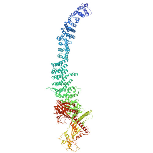
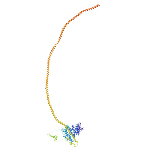
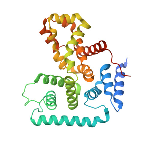
Structure of the human TSC:WIPI3 lysosomal recruitment complex.

Bayly-Jones, C.Lupton, C.J.D'Andrea, L.Chang, Y.G.Jones, G.D.Steele, J.R.Venugopal, H.Schittenhelm, R.B.Halls, M.L.Ellisdon, A.M.

(2024) Sci Adv 10: eadr5807-eadr5807

  • DOI: https://doi.org/10.1126/sciadv.adr5807
  • Primary Citation of Related Structures:  
    9C9I, 9CE3

  • PubMed Abstract: 

    Tuberous sclerosis complex (TSC) is targeted to the lysosomal membrane, where it hydrolyzes RAS homolog-mTORC1 binding (RHEB) from its GTP-bound to GDP-bound state, inhibiting mechanistic target of rapamycin complex 1 (mTORC1). Loss-of-function mutations in TSC cause TSC disease, marked by excessive tumor growth. Here, we overcome a high degree of continuous conformational heterogeneity to determine the 2.8-Å cryo-electron microscopy (cryo-EM) structure of the complete human TSC in complex with the lysosomal recruitment factor WD repeat domain phosphoinositide-interacting protein 3 (WIPI3). We discover a previously undetected amino-terminal TSC1 HEAT repeat dimer that clamps onto a single TSC wing and forms a phosphatidylinositol phosphate (PIP)-binding pocket, which specifically binds monophosphorylated PIPs. These structural advances provide a model by which WIPI3 and PIP-signaling networks coordinate to recruit TSC to the lysosomal membrane to inhibit mTORC1. The high-resolution TSC structure reveals previously unrecognized mutational hotspots and uncovers crucial insights into the mechanisms of TSC dysregulation in disease.


  • Organizational Affiliation
    • Cancer Program, Biomedicine Discovery Institute, Monash University, Clayton, VIC 3800, Australia.

Macromolecules
Find similar proteins by:  (by identity cutoff)  |  3D Structure
Entity ID: 1
MoleculeChains Sequence LengthOrganismDetailsImage
Isoform 4 of Tuberin
A, B
1,792Homo sapiensMutation(s): 0 
Gene Names: TSC2TSC4
UniProt & NIH Common Fund Data Resources
Find proteins for P49815 (Homo sapiens)
Explore P49815 
Go to UniProtKB:  P49815
PHAROS:  P49815
GTEx:  ENSG00000103197 
Entity Groups  
Sequence Clusters30% Identity50% Identity70% Identity90% Identity95% Identity100% Identity
UniProt GroupP49815
Sequence Annotations
Expand
  • Reference Sequence
Find similar proteins by:  (by identity cutoff)  |  3D Structure
Entity ID: 2
MoleculeChains Sequence LengthOrganismDetailsImage
Hamartin
C, D
1,191Homo sapiensMutation(s): 0 
Gene Names: TSC1KIAA0243TSC
UniProt & NIH Common Fund Data Resources
Find proteins for Q92574 (Homo sapiens)
Explore Q92574 
Go to UniProtKB:  Q92574
PHAROS:  Q92574
GTEx:  ENSG00000165699 
Entity Groups  
Sequence Clusters30% Identity50% Identity70% Identity90% Identity95% Identity100% Identity
UniProt GroupQ92574
Sequence Annotations
Expand
  • Reference Sequence
Find similar proteins by:  (by identity cutoff)  |  3D Structure
Entity ID: 3
MoleculeChains Sequence LengthOrganismDetailsImage
TBC1 domain family member 7301Homo sapiensMutation(s): 0 
Gene Names: TBC1D7TBC7HSPC239
UniProt & NIH Common Fund Data Resources
Find proteins for Q9P0N9 (Homo sapiens)
Explore Q9P0N9 
Go to UniProtKB:  Q9P0N9
PHAROS:  Q9P0N9
GTEx:  ENSG00000145979 
Entity Groups  
Sequence Clusters30% Identity50% Identity70% Identity90% Identity95% Identity100% Identity
UniProt GroupQ9P0N9
Sequence Annotations
Expand
  • Reference Sequence
Find similar proteins by:  (by identity cutoff)  |  3D Structure
Entity ID: 4
MoleculeChains Sequence LengthOrganismDetailsImage
WD repeat domain phosphoinositide-interacting protein 3316Homo sapiensMutation(s): 0 
Gene Names: WDR45BWDR45LWIPI3
UniProt & NIH Common Fund Data Resources
Find proteins for Q5MNZ6 (Homo sapiens)
Explore Q5MNZ6 
Go to UniProtKB:  Q5MNZ6
PHAROS:  Q5MNZ6
GTEx:  ENSG00000141580 
Entity Groups  
Sequence Clusters30% Identity50% Identity70% Identity90% Identity95% Identity100% Identity
UniProt GroupQ5MNZ6
Sequence Annotations
Expand
  • Reference Sequence

Find similar proteins by:  Sequence   |   3D Structure  

Entity ID: 5
MoleculeChains Sequence LengthOrganismDetailsImage
Unknown fragment
G, H
12Homo sapiensMutation(s): 0 
Sequence Annotations
Expand
  • Reference Sequence
Experimental Data & Validation

Experimental Data

  • Method: ELECTRON MICROSCOPY
  • Resolution: 2.90 Å
  • Aggregation State: PARTICLE 
  • Reconstruction Method: SINGLE PARTICLE 
EM Software:
TaskSoftware PackageVersion
MODEL REFINEMENTPHENIX
MODEL REFINEMENTCoot
MODEL REFINEMENTISOLDE

Structure Validation

View Full Validation Report


Entry History & Funding Information

Deposition Data


Funding OrganizationLocationGrant Number
Department of Defense (DOD, United States)United StatesW81XWH-19-1-0182
Australian Research Council (ARC)AustraliaDE240100992
Australian Research Council (ARC)AustraliaLE170100016

Revision History  (Full details and data files)

  • Version 1.0: 2024-12-04
    Type: Initial release