9CGI | pdb_00009cgi

Cryo-EM structure of the Nipah Virus polymerase (L) protein in complex with the tetrameric phosphoprotein (P)


Experimental Data Snapshot

  • Method: ELECTRON MICROSCOPY
  • Resolution: 2.92 Å
  • Aggregation State: PARTICLE 
  • Reconstruction Method: SINGLE PARTICLE 

wwPDB Validation   3D Report Full Report


This is version 1.1 of the entry. See complete history


Literature

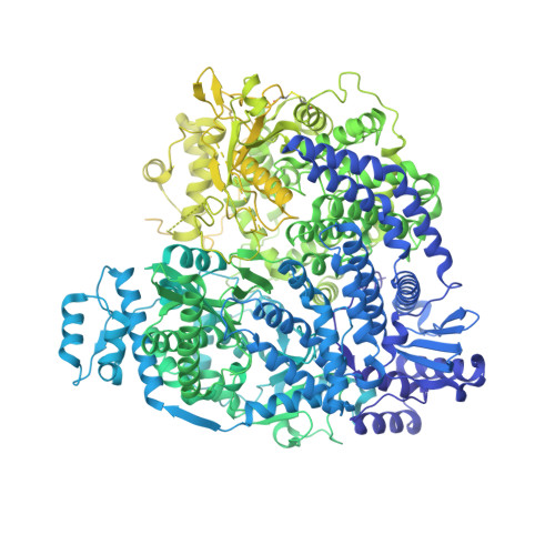
Structure of the Nipah virus polymerase phosphoprotein complex.

Yang, G.Wang, D.Liu, B.

(2024) Nat Commun 15: 8673-8673

  • DOI: https://doi.org/10.1038/s41467-024-52701-y
  • Primary Citation of Related Structures:  
    9CGI

  • PubMed Abstract: 

    The Nipah virus (NiV), a member of the Paramyxoviridae family, is notorious for its high fatality rate in humans. The RNA polymerase machinery of NiV, comprising the large protein L and the phosphoprotein P, is essential for viral replication. This study presents the 2.9-Å cryo-electron microscopy structure of the NiV L-P complex, shedding light on its assembly and functionality. The structure not only demonstrates the molecular details of the conserved N-terminal domain, RNA-dependent RNA polymerase (RdRp), and GDP polyribonucleotidyltransferase of the L protein, but also the intact central oligomerization domain and the C-terminal X domain of the P protein. The P protein interacts extensively with the L protein, forming an antiparallel β-sheet among the P protomers and with the fingers subdomain of RdRp. The flexible linker domain of one P promoter extends its contact with the fingers subdomain to reach near the nascent RNA exit, highlighting the distinct characteristic of the NiV L-P interface. This distinctive tetrameric organization of the P protein and its interaction with the L protein provide crucial molecular insights into the replication and transcription mechanisms of NiV polymerase, ultimately contributing to the development of effective treatments and preventive measures against this Paramyxoviridae family deadly pathogen.


  • Organizational Affiliation
    • Section of Transcription & Gene Regulation, The Hormel Institute, University of Minnesota, Austin, MN, USA.

Macromolecules
Find similar proteins by:  (by identity cutoff)  |  3D Structure
Entity ID: 1
MoleculeChains Sequence LengthOrganismDetailsImage
RNA-directed RNA polymerase L2,244Henipavirus nipahenseMutation(s): 0 
EC: 2.7.7.48 (PDB Primary Data), 3.6.1 (PDB Primary Data), 2.7.7.88 (PDB Primary Data), 2.1.1.375 (PDB Primary Data)
UniProt
Find proteins for Q997F0 (Nipah virus)
Explore Q997F0 
Go to UniProtKB:  Q997F0
Entity Groups  
Sequence Clusters30% Identity50% Identity70% Identity90% Identity95% Identity100% Identity
UniProt GroupQ997F0
Sequence Annotations
Expand
  • Reference Sequence
Find similar proteins by:  (by identity cutoff)  |  3D Structure
Entity ID: 2
MoleculeChains Sequence LengthOrganismDetailsImage
Phosphoprotein
B, C, D, E
709Henipavirus nipahenseMutation(s): 0 
Gene Names: P/V/C
UniProt
Find proteins for Q9IK91 (Nipah virus)
Explore Q9IK91 
Go to UniProtKB:  Q9IK91
Entity Groups  
Sequence Clusters30% Identity50% Identity70% Identity90% Identity95% Identity100% Identity
UniProt GroupQ9IK91
Sequence Annotations
Expand
  • Reference Sequence
Experimental Data & Validation

Experimental Data

  • Method: ELECTRON MICROSCOPY
  • Resolution: 2.92 Å
  • Aggregation State: PARTICLE 
  • Reconstruction Method: SINGLE PARTICLE 
EM Software:
TaskSoftware PackageVersion
MODEL REFINEMENTPHENIX1.21_5207:

Structure Validation

View Full Validation Report


Entry History & Funding Information

Deposition Data


Funding OrganizationLocationGrant Number
Not funded--

Revision History  (Full details and data files)

  • Version 1.0: 2024-09-18
    Type: Initial release
  • Version 1.1: 2024-10-23
    Changes: Data collection, Database references, Structure summary