9CR8

SapNP reconstituted human ABCB1


Experimental Data Snapshot

  • Method: ELECTRON MICROSCOPY
  • Resolution: 3.80 Å
  • Aggregation State: PARTICLE 
  • Reconstruction Method: SINGLE PARTICLE 

wwPDB Validation   3D Report Full Report


This is version 1.0 of the entry. See complete history


Literature

Structural insights into binding-site access and ligand recognition by human ABCB1.

Kurre, D.Dang, P.X.Le, L.T.M.Gadkari, V.V.Alam, A.

(2025) EMBO J 

  • DOI: https://doi.org/10.1038/s44318-025-00361-z
  • Primary Citation of Related Structures:  
    9CR8, 9CTC, 9CTF, 9CTG

  • PubMed Abstract: 

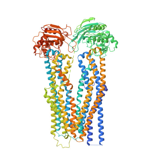
    ABCB1 is a broad-spectrum efflux pump central to cellular drug handling and multidrug resistance in humans. However, how it is able to recognize and transport a wide range of diverse substrates remains poorly understood. Here we present cryo-EM structures of lipid-embedded human ABCB1 in conformationally distinct apo-, substrate-bound, inhibitor-bound, and nucleotide-trapped states at 3.4-3.9 Å resolution, in the absence of stabilizing antibodies or mutations. The substrate-binding site is located within one half of the molecule and, in the apo state, is obstructed by the transmembrane helix (TM) 4. Substrate and inhibitor binding are distinguished by major TM rearrangements and their ligand binding chemistry, with TM4 playing a central role in all conformational transitions. Furthermore, our data identify secondary structure-breaking residues that impart localized TM flexibility and asymmetry between the two transmembrane domains. The resulting structural changes and lipid interactions that are induced by substrate and inhibitor binding can predict substrate-binding profiles and may direct ABCB1 inhibitor design.


  • Organizational Affiliation

    The Hormel Institute, University of Minnesota, Austin, MN, 55912, USA.


Macromolecules
Find similar proteins by:  (by identity cutoff)  |  3D Structure
Entity ID: 1
MoleculeChains Sequence LengthOrganismDetailsImage
ATP-dependent translocase ABCB11,280Homo sapiensMutation(s): 0 
Gene Names: ABCB1MDR1PGY1
EC: 7.6.2.2 (PDB Primary Data), 7.6.2.1 (PDB Primary Data)
Membrane Entity: Yes 
UniProt & NIH Common Fund Data Resources
Find proteins for P08183 (Homo sapiens)
Explore P08183 
Go to UniProtKB:  P08183
PHAROS:  P08183
GTEx:  ENSG00000085563 
Entity Groups  
Sequence Clusters30% Identity50% Identity70% Identity90% Identity95% Identity100% Identity
UniProt GroupP08183
Sequence Annotations
Expand
  • Reference Sequence
Small Molecules
Ligands 1 Unique
IDChains Name / Formula / InChI Key2D Diagram3D Interactions
UPL (Subject of Investigation/LOI)
Query on UPL

Download Ideal Coordinates CCD File 
B [auth A]
C [auth A]
D [auth A]
E [auth A]
F [auth A]
B [auth A],
C [auth A],
D [auth A],
E [auth A],
F [auth A],
G [auth A],
H [auth A],
I [auth A],
J [auth A],
K [auth A],
L [auth A],
M [auth A],
N [auth A],
O [auth A],
P [auth A]
UNKNOWN BRANCHED FRAGMENT OF PHOSPHOLIPID
C34 H70
QPCSNFPFMCVVAI-UHFFFAOYSA-N
Experimental Data & Validation

Experimental Data

  • Method: ELECTRON MICROSCOPY
  • Resolution: 3.80 Å
  • Aggregation State: PARTICLE 
  • Reconstruction Method: SINGLE PARTICLE 
EM Software:
TaskSoftware PackageVersion
RECONSTRUCTIONRELION

Structure Validation

View Full Validation Report


Entry History & Funding Information

Deposition Data


Funding OrganizationLocationGrant Number
National Institutes of Health/National Institute of General Medical Sciences (NIH/NIGMS)United States1R01GM146906

Revision History  (Full details and data files)

  • Version 1.0: 2025-01-22
    Type: Initial release