9CXM | pdb_00009cxm

Crystal structure of Human FN3K bound with ATP and DMF


Experimental Data Snapshot

  • Method: X-RAY DIFFRACTION
  • Resolution: 1.76 Å
  • R-Value Free: 
    0.208 (Depositor), 0.210 (DCC) 
  • R-Value Work: 
    0.176 (Depositor), 0.180 (DCC) 
  • R-Value Observed: 
    0.178 (Depositor) 

Starting Model: experimental
View more details

wwPDB Validation   3D Report Full Report


Ligand Structure Quality Assessment 


This is version 1.1 of the entry. See complete history


Literature

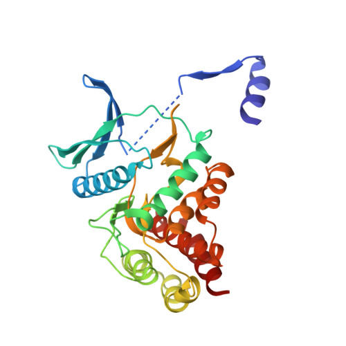
The molecular basis of Human FN3K mediated phosphorylation of glycated substrates.

Garg, A.On, K.F.Xiao, Y.Elkayam, E.Cifani, P.David, Y.Joshua-Tor, L.

(2025) Nat Commun 16: 941-941

  • DOI: https://doi.org/10.1038/s41467-025-56207-z
  • Primary Citation of Related Structures:  
    9CX8, 9CXM, 9CXN, 9CXO, 9CXV, 9CXW

  • PubMed Abstract: 

    Glycation, a non-enzymatic post-translational modification occurring on proteins, can be actively reversed via site-specific phosphorylation of the fructose-lysine moiety by FN3K kinase, to impact the cellular function of the target protein. A regulatory axis between FN3K and glycated protein targets has been associated with conditions like diabetes and cancer. However, the molecular basis of this relationship has not been explored so far. Here, we determined a series of crystal structures of HsFN3K in the apo-state, and in complex with different nucleotide analogs together with a sugar substrate mimic to reveal the features important for its kinase activity and substrate recognition. Additionally, the dynamics in sugar substrate binding during the kinase catalytic cycle provide important mechanistic insights into HsFN3K function. Our structural work provides the molecular basis for rational small molecule design targeting FN3K.


  • Organizational Affiliation
    • Cold Spring Harbor Laboratory, One Bungtown Road, Cold Spring Harbor, New York, 11724, USA.

Macromolecules
Find similar proteins by:  (by identity cutoff)  |  3D Structure
Entity ID: 1
MoleculeChains Sequence LengthOrganismDetailsImage
Fructosamine-3-kinase
A, B
291Homo sapiensMutation(s): 0 
Gene Names: FN3K
EC: 2.7.1.171 (PDB Primary Data), 2.7.1.172 (PDB Primary Data)
UniProt & NIH Common Fund Data Resources
Find proteins for Q9H479 (Homo sapiens)
Explore Q9H479 
Go to UniProtKB:  Q9H479
PHAROS:  Q9H479
GTEx:  ENSG00000167363 
Entity Groups  
Sequence Clusters30% Identity50% Identity70% Identity90% Identity95% Identity100% Identity
UniProt GroupQ9H479
Sequence Annotations
Expand
  • Reference Sequence
Small Molecules
Ligands 5 Unique
IDChains Name / Formula / InChI Key2D Diagram3D Interactions
ATP (Subject of Investigation/LOI)
Query on ATP

Download Ideal Coordinates CCD File 
Q [auth B]ADENOSINE-5'-TRIPHOSPHATE
C10 H16 N5 O13 P3
ZKHQWZAMYRWXGA-KQYNXXCUSA-N
ADP (Subject of Investigation/LOI)
Query on ADP

Download Ideal Coordinates CCD File 
J [auth A]ADENOSINE-5'-DIPHOSPHATE
C10 H15 N5 O10 P2
XTWYTFMLZFPYCI-KQYNXXCUSA-N
A1A0O (Subject of Investigation/LOI)
Query on A1A0O

Download Ideal Coordinates CCD File 
R [auth B]1-deoxy-1-(morpholin-4-yl)-D-fructose
C10 H19 N O6
JPANQHVKLYKLEB-OPRDCNLKSA-N
SO4
Query on SO4

Download Ideal Coordinates CCD File 
C [auth A],
N [auth B],
O [auth B]
SULFATE ION
O4 S
QAOWNCQODCNURD-UHFFFAOYSA-L
EDO
Query on EDO

Download Ideal Coordinates CCD File 
D [auth A]
E [auth A]
F [auth A]
G [auth A]
H [auth A]
D [auth A],
E [auth A],
F [auth A],
G [auth A],
H [auth A],
I [auth A],
K [auth A],
L [auth A],
M [auth A],
P [auth B],
S [auth B],
T [auth B],
U [auth B],
V [auth B],
W [auth B]
1,2-ETHANEDIOL
C2 H6 O2
LYCAIKOWRPUZTN-UHFFFAOYSA-N
Experimental Data & Validation

Experimental Data

  • Method: X-RAY DIFFRACTION
  • Resolution: 1.76 Å
  • R-Value Free:  0.208 (Depositor), 0.210 (DCC) 
  • R-Value Work:  0.176 (Depositor), 0.180 (DCC) 
  • R-Value Observed: 0.178 (Depositor) 
Space Group: P 21 21 21
Unit Cell:
Length ( Å )Angle ( ˚ )
a = 52.932α = 90
b = 112.275β = 90
c = 132.796γ = 90
Software Package:
Software NamePurpose
PHENIXrefinement
Cootmodel building
RAPDdata processing
PHASERphasing
RAPDdata scaling
RAPDdata reduction

Structure Validation

View Full Validation Report


Ligand Structure Quality Assessment 


Entry History & Funding Information

Deposition Data


Funding OrganizationLocationGrant Number
Howard Hughes Medical Institute (HHMI)United States--

Revision History  (Full details and data files)

  • Version 1.0: 2024-12-18
    Type: Initial release
  • Version 1.1: 2025-02-05
    Changes: Database references