Isoleucine Binding and Regulation of Escherichia coli and Staphylococcus aureus Threonine Dehydratase (IlvA).
Yun, M.K., Subramanian, C., Miller, K., Jackson, P., Radka, C.D., Rock, C.O.(2025) Biochemistry 64: 2793-2810
- PubMed: 40494512
- DOI: https://doi.org/10.1021/acs.biochem.5c00168
- Primary Citation of Related Structures:
9D2Q, 9D2R, 9D2S, 9D2T - PubMed Abstract:
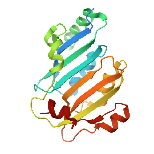
In Staphylococcus aureus , the branched-chain amino acid biosynthetic pathway provides essential intermediates for membrane biosynthesis. Threonine deaminase (IlvA) is the first enzyme in the pathway, and isoleucine feedback regulates the enzyme in Escherichia coli . These studies on E. coli IlvA (EcIlvA) introduced the concept of allosteric regulation. To investigate the regulation of S. aureus IlvA (SaIlvA), we first conducted additional studies on EcIlvA. The previously determined crystal structure of EcIlvA revealed a tetrameric assembly of protomers, each with catalytic and regulatory domains, but the structural basis of isoleucine regulation was not characterized. Here, we present the crystal structure of the EcIlvA regulatory domain bound to isoleucine, which reveals the isoleucine binding site and conformational changes that initiate at Phe352 and propagate 23 Å across the domain. This suggests an allosteric pathway that extends to the active site of the adjacent protomer, mediating regulation across the protomer-protomer interface. The EcIlvA(F352A) mutant binds isoleucine but is feedback-resistant due to the absence of the initiating Phe352. In contrast, SaIlvA is not feedback-regulated by isoleucine and does not bind it. The structure of the SaIlvA regulatory domain reveals a different organization that lacks the isoleucine binding site. Other potential allosteric inhibitors of SaIlvA, including phospholipid intermediates, do not affect enzyme activity. We propose that the absence of feedback inhibition in SaIlvA is due to its role in membrane biosynthesis. These findings enhance our understanding of IlvA's allosteric regulation and offer opportunities for engineering feedback-resistant IlvA variants for biotechnological use.
Organizational Affiliation:
Department of Host-Microbe Interactions, St. Jude Children's Research Hospital, Memphis, Tennessee 38105, United States.