9DBN | pdb_00009dbn

Tarantula venom peptide Protoxin-I bound to full-length human voltage-gated sodium channel 1.8 (NaV1.8)


Experimental Data Snapshot

  • Method: ELECTRON MICROSCOPY
  • Resolution: 2.76 Å
  • Aggregation State: PARTICLE 
  • Reconstruction Method: SINGLE PARTICLE 

Starting Models: experimental
View more details

wwPDB Validation   3D Report Full Report


This is version 1.0 of the entry. See complete history


Literature

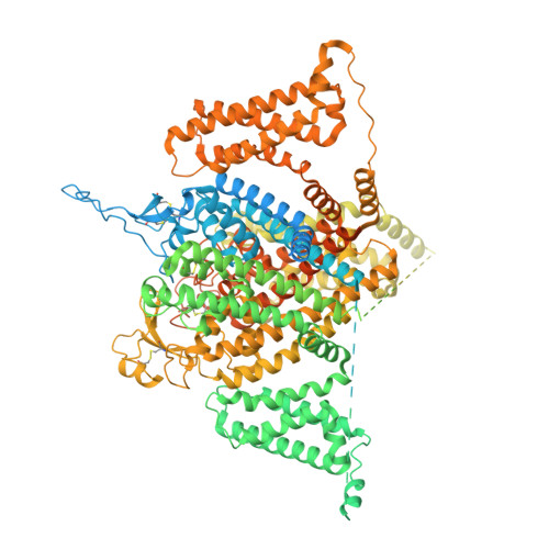
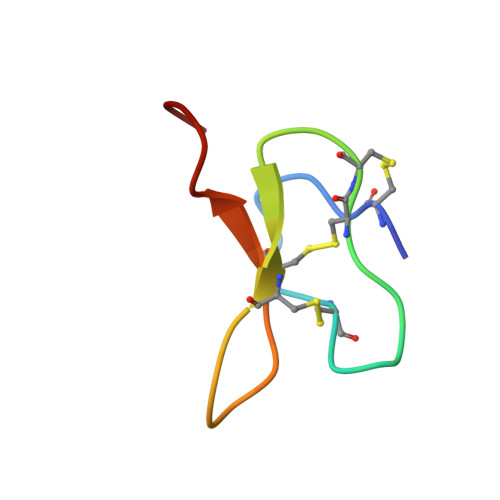
Structural basis of inhibition of human Na V 1.8 by the tarantula venom peptide Protoxin-I.

Neumann, B.McCarthy, S.Gonen, S.

(2025) Nat Commun 16: 1459-1459

  • DOI: https://doi.org/10.1038/s41467-024-55764-z
  • Primary Citation of Related Structures:  
    9DBK, 9DBL, 9DBM, 9DBN

  • PubMed Abstract: 

    Voltage-gated sodium channels (Na V s) selectively permit diffusion of sodium ions across the cell membrane and, in excitable cells, are responsible for propagating action potentials. One of the nine human Na V isoforms, Na V 1.8, is a promising target for analgesics, and selective inhibitors are of interest as therapeutics. One such inhibitor, the gating-modifier peptide Protoxin-I derived from tarantula venom, blocks channel opening by shifting the activation voltage threshold to more depolarized potentials, but the structural basis for this inhibition has not previously been determined. Using monolayer graphene grids, we report the cryogenic electron microscopy structures of full-length human apo-Na V 1.8 and the Protoxin-I-bound complex at 3.1 Å and 2.8 Å resolution, respectively. The apo structure shows an unexpected movement of the Domain I S4-S5 helix, and VSD I was unresolvable. We find that Protoxin-I binds to and displaces the VSD II S3-S4 linker, hindering translocation of the S4 II helix during activation.


  • Organizational Affiliation
    • Department of Molecular Biology and Biochemistry, University of California Irvine, Irvine, CA, USA.

Macromolecules
Find similar proteins by:  (by identity cutoff)  |  3D Structure
Entity ID: 1
MoleculeChains Sequence LengthOrganismDetailsImage
Sodium channel protein type 10 subunit alpha2,001Homo sapiensMutation(s): 1 
Gene Names: SCN10A
Membrane Entity: Yes 
UniProt & NIH Common Fund Data Resources
Find proteins for Q9Y5Y9 (Homo sapiens)
Explore Q9Y5Y9 
Go to UniProtKB:  Q9Y5Y9
PHAROS:  Q9Y5Y9
GTEx:  ENSG00000185313 
Entity Groups  
Sequence Clusters30% Identity50% Identity70% Identity90% Identity95% Identity100% Identity
UniProt GroupQ9Y5Y9
Glycosylation
Glycosylation Sites: 5Go to GlyGen: Q9Y5Y9-1
Sequence Annotations
Expand
  • Reference Sequence
Find similar proteins by:  (by identity cutoff)  |  3D Structure
Entity ID: 2
MoleculeChains Sequence LengthOrganismDetailsImage
Beta/omega-theraphotoxin-Tp1a35Thrixopelma pruriensMutation(s): 0 
UniProt
Find proteins for P83480 (Thrixopelma pruriens)
Explore P83480 
Go to UniProtKB:  P83480
Entity Groups  
Sequence Clusters30% Identity50% Identity70% Identity90% Identity95% Identity100% Identity
UniProt GroupP83480
Sequence Annotations
Expand
  • Reference Sequence
Oligosaccharides

Help

Entity ID: 3
MoleculeChains Length2D Diagram Glycosylation3D Interactions
beta-D-mannopyranose-(1-4)-2-acetamido-2-deoxy-beta-D-glucopyranose-(1-4)-2-acetamido-2-deoxy-beta-D-glucopyranose
C, D, E, F, G
3N-Glycosylation
Glycosylation Resources
GlyTouCan:  G15407YE
GlyCosmos:  G15407YE
GlyGen:  G15407YE
Small Molecules
Ligands 4 Unique
IDChains Name / Formula / InChI Key2D Diagram3D Interactions
P5S
Query on P5S

Download Ideal Coordinates CCD File 
S [auth A],
T [auth A]
O-[(R)-{[(2R)-2,3-bis(octadecanoyloxy)propyl]oxy}(hydroxy)phosphoryl]-L-serine
C42 H82 N O10 P
TZCPCKNHXULUIY-RGULYWFUSA-N
PCW
Query on PCW

Download Ideal Coordinates CCD File 
K [auth A],
N [auth A],
O [auth A]
1,2-DIOLEOYL-SN-GLYCERO-3-PHOSPHOCHOLINE
C44 H85 N O8 P
SNKAWJBJQDLSFF-NVKMUCNASA-O
LPE
Query on LPE

Download Ideal Coordinates CCD File 
L [auth A],
M [auth A],
P [auth A],
Q [auth A],
R [auth A]
1-O-OCTADECYL-SN-GLYCERO-3-PHOSPHOCHOLINE
C26 H57 N O6 P
XKBJVQHMEXMFDZ-AREMUKBSSA-O
CLR
Query on CLR

Download Ideal Coordinates CCD File 
H [auth A],
I [auth A],
J [auth A],
U [auth A]
CHOLESTEROL
C27 H46 O
HVYWMOMLDIMFJA-DPAQBDIFSA-N
Experimental Data & Validation

Experimental Data

  • Method: ELECTRON MICROSCOPY
  • Resolution: 2.76 Å
  • Aggregation State: PARTICLE 
  • Reconstruction Method: SINGLE PARTICLE 
EM Software:
TaskSoftware PackageVersion
RECONSTRUCTIONcryoSPARC4.4
MODEL REFINEMENTPHENIX
MODEL REFINEMENTCoot

Structure Validation

View Full Validation Report


Entry History & Funding Information

Deposition Data


Funding OrganizationLocationGrant Number
Department of Defense (DOD, United States)United StatesHDTRA1-21-1-0004
National Institutes of Health/National Institute of General Medical Sciences (NIH/NIGMS)United StatesR35-GM142797

Revision History  (Full details and data files)

  • Version 1.0: 2025-02-19
    Type: Initial release