9DC6 | pdb_00009dc6

Structure of J-PKAc chimera in complex with Aplithianine e1


Experimental Data Snapshot

  • Method: X-RAY DIFFRACTION
  • Resolution: 2.70 Å
  • R-Value Free: 
    0.311 (Depositor), 0.307 (DCC) 
  • R-Value Work: 
    0.232 (Depositor), 0.229 (DCC) 
  • R-Value Observed: 
    0.237 (Depositor) 

Starting Model: experimental
View more details

wwPDB Validation   3D Report Full Report


Ligand Structure Quality Assessment 


This is version 1.0 of the entry. See complete history


Literature

Chemical Evolution of Aplithianine Class of Serine/Threonine Kinase Inhibitors.

Du, L.Wilson, B.A.P.Moore, W.J.Dalilian, M.Shenoy, S.R.Li, N.Martinez Fiesco, J.A.Hwang, J.Y.Smith, E.A.Wamiru, A.Goncharova, E.I.Alvarez de la Cruz, A.Pagadala, K.Piswa, H.K.Patteti, V.Jampana, V.P.Manepalli, P.Nimmala, R.Marri, N.R.Gunuguntla, M.Reddy, J.J.Schneekloth Jr., J.S.Zhang, P.O'Keefe, B.R.

(2025) J Med Chem 68: 12756-12785

  • DOI: https://doi.org/10.1021/acs.jmedchem.5c00649
  • Primary Citation of Related Structures:  
    9DC6, 9DCD

  • PubMed Abstract: 

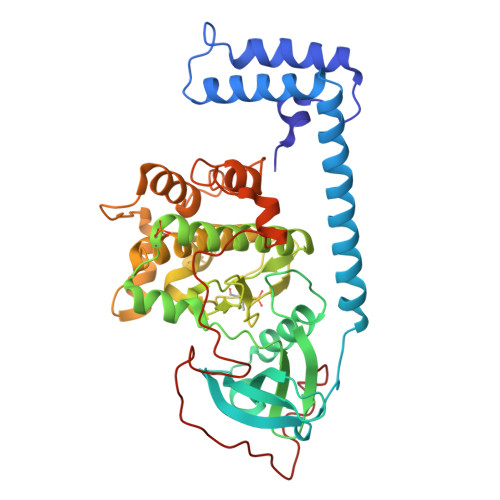
    Chimeric kinase J-PKAcα represents a potential therapeutic target for fibrolamellar hepatocellular carcinoma (FLHCC). Structure-based design and screening were applied to improve the potency of the marine-derived kinase inhibitor aplithianine A targeting J-PKAcα. Three classes of aplithianines (I, II, and III) including >150 analogs were synthesized, significantly improving biochemical IC 50 values to the low nanomolar range. X-ray diffraction experiments confirmed that the class II aplithianines adopted a novel binding mode to J-PKAcα by interacting with the DFG residue Asp239. The kinase selectivity profiles were assessed by kinome profiling. In vitro profiles of selected class II analogs were evaluated to determine compound solubility, protein binding, permeability, metabolism, and hERG binding. Selected aplithianine analogs inhibited intracellular phosphorylation of the peptide substrate CREB following stimulation of the J-PKAcα fusion kinase in NIH/3T3 cells and exhibited antiproliferative/cytotoxic activities against select cancer cell lines from the NCI-60 cell panel at nanomolar concentrations.


  • Organizational Affiliation
    • Molecular Targets Program, Center for Cancer Research, National Cancer Institute, Frederick, Maryland 21702, United States.

Macromolecules
Find similar proteins by:  (by identity cutoff)  |  3D Structure
Entity ID: 1
MoleculeChains Sequence LengthOrganismDetailsImage
cAMP-dependent protein kinase catalytic subunit alpha
A, B
405Homo sapiensMutation(s): 0 
Gene Names: PRKACAPKACA
EC: 2.7.11.11
UniProt & NIH Common Fund Data Resources
Find proteins for P17612 (Homo sapiens)
Explore P17612 
Go to UniProtKB:  P17612
PHAROS:  P17612
GTEx:  ENSG00000072062 
Entity Groups  
Sequence Clusters30% Identity50% Identity70% Identity90% Identity95% Identity100% Identity
UniProt GroupP17612
Sequence Annotations
Expand
  • Reference Sequence

Find similar proteins by:  Sequence   |   3D Structure  

Entity ID: 2
MoleculeChains Sequence LengthOrganismDetailsImage
cAMP-dependent protein kinase inhibitor alphaC [auth I],
D [auth J]
20Homo sapiensMutation(s): 0 
UniProt & NIH Common Fund Data Resources
Find proteins for P61925 (Homo sapiens)
Explore P61925 
Go to UniProtKB:  P61925
PHAROS:  P61925
GTEx:  ENSG00000171033 
Entity Groups  
Sequence Clusters30% Identity50% Identity70% Identity90% Identity95% Identity100% Identity
UniProt GroupP61925
Sequence Annotations
Expand
  • Reference Sequence
Small Molecules
Ligands 1 Unique
IDChains Name / Formula / InChI Key2D Diagram3D Interactions
A1A3I (Subject of Investigation/LOI)
Query on A1A3I

Download Ideal Coordinates CCD File 
E [auth A],
F [auth B]
N-(2-aminoethyl)-4-(7H-pyrrolo[2,3-d]pyrimidin-4-yl)-3,4-dihydro-2H-1,4-thiazine-6-carboxamide
C13 H16 N6 O S
CTPKBBPXVZYOPM-UHFFFAOYSA-N
Modified Residues  2 Unique
IDChains TypeFormula2D DiagramParent
SEP
Query on SEP
A, B
L-PEPTIDE LINKINGC3 H8 N O6 PSER
TPO
Query on TPO
A, B
L-PEPTIDE LINKINGC4 H10 N O6 PTHR
Experimental Data & Validation

Experimental Data

  • Method: X-RAY DIFFRACTION
  • Resolution: 2.70 Å
  • R-Value Free:  0.311 (Depositor), 0.307 (DCC) 
  • R-Value Work:  0.232 (Depositor), 0.229 (DCC) 
  • R-Value Observed: 0.237 (Depositor) 
Space Group: P 21 21 21
Unit Cell:
Length ( Å )Angle ( ˚ )
a = 60.758α = 90
b = 124.934β = 90
c = 129.302γ = 90
Software Package:
Software NamePurpose
PHENIXrefinement
XDSdata reduction
XDSdata scaling
PHASERphasing

Structure Validation

View Full Validation Report


Ligand Structure Quality Assessment 


Entry History & Funding Information

Deposition Data


Funding OrganizationLocationGrant Number
National Institutes of Health/National Cancer Institute (NIH/NCI)United States--

Revision History  (Full details and data files)

  • Version 1.0: 2025-07-09
    Type: Initial release