9DEV | pdb_00009dev

PARP4 BRCT domain


Experimental Data Snapshot

  • Method: X-RAY DIFFRACTION
  • Resolution: 1.75 Å
  • R-Value Free: 
    0.188 (Depositor), 0.200 (DCC) 
  • R-Value Work: 
    0.175 (Depositor), 0.186 (DCC) 
  • R-Value Observed: 
    0.175 (Depositor) 

Starting Model: in silico
View more details

wwPDB Validation   3D Report Full Report


Ligand Structure Quality Assessment 


This is version 1.2 of the entry. See complete history


Literature

Crystal structure and mutagenesis of a nucleic acid-binding BRCT domain in human PARP4.

Frigon, L.Pascal, J.M.

(2025) J Biological Chem 301: 110277-110277

  • DOI: https://doi.org/10.1016/j.jbc.2025.110277
  • Primary Citation of Related Structures:  
    9DEV, 9DFO, 9DFP, 9DFQ, 9DFR

  • PubMed Abstract: 

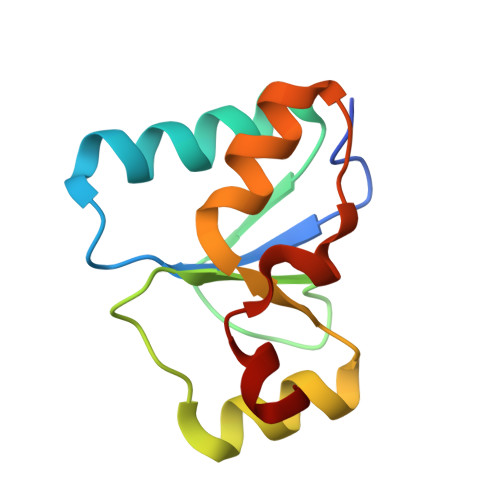
    PARP4 is an ADP-ribosyltransferase typically associated with the cytoplasmic vault organelle. PARP4 has a distinct domain composition relative to other PARP enzymes; however, the N-terminal region of PARP4 is homologous to a collection of domains found in PARP1, a regulator of multiple nuclear processes including the cellular response to DNA damage. The N-terminal region of PARP4 interacts in vitro with nucleic acid, in particular a non-coding RNA associated with vault particles, and a BRCT domain is implicated in this interaction. Here we report the X-ray structure of the BRCT domain of PARP4 and structure-based mutagenesis that interrogates the nucleic acid binding activity using vault RNA. The isolated BRCT domain is capable of mediating interaction with vault RNA, and we identified four BRCT mutants that disrupt vault RNA interaction to varying degrees. X-ray structures of the BRCT mutants indicate that perturbations to an electropositive region of the BRCT surface underlie the loss of nucleic acid binding. Comparison to other nucleic acid-binding BRCT domains highlights distinct features of the PARP4 BRCT structure. The study presents the experimental structure of the PARP4 BRCT domain, establishes this domain as nucleic acid-binding module, and provides PARP4 BRCT mutants that can be used to investigate PARP4 cellular functions.


  • Organizational Affiliation
    • Department of Biochemistry and Molecular Medicine, Université de Montréal, Montréal, Qc H3T 1J4, Canada.

Macromolecules
Find similar proteins by:  (by identity cutoff)  |  3D Structure
Entity ID: 1
MoleculeChains Sequence LengthOrganismDetailsImage
Protein mono-ADP-ribosyltransferase PARP4100Homo sapiensMutation(s): 0 
Gene Names: PARP4ADPRTL1KIAA0177PARPL
EC: 2.4.2
UniProt & NIH Common Fund Data Resources
Find proteins for Q9UKK3 (Homo sapiens)
Explore Q9UKK3 
Go to UniProtKB:  Q9UKK3
PHAROS:  Q9UKK3
GTEx:  ENSG00000102699 
Entity Groups  
Sequence Clusters30% Identity50% Identity70% Identity90% Identity95% Identity100% Identity
UniProt GroupQ9UKK3
Sequence Annotations
Expand
  • Reference Sequence
Small Molecules
Experimental Data & Validation

Experimental Data

  • Method: X-RAY DIFFRACTION
  • Resolution: 1.75 Å
  • R-Value Free:  0.188 (Depositor), 0.200 (DCC) 
  • R-Value Work:  0.175 (Depositor), 0.186 (DCC) 
  • R-Value Observed: 0.175 (Depositor) 
Space Group: F 2 3
Unit Cell:
Length ( Å )Angle ( ˚ )
a = 114.428α = 90
b = 114.428β = 90
c = 114.428γ = 90
Software Package:
Software NamePurpose
REFMACrefinement
Aimlessdata scaling
PHENIXphasing
XDSdata reduction

Structure Validation

View Full Validation Report


Ligand Structure Quality Assessment 


Entry History & Funding Information

Deposition Data


Funding OrganizationLocationGrant Number
Canadian Institutes of Health Research (CIHR)CanadaPJT153295

Revision History  (Full details and data files)

  • Version 1.0: 2025-05-28
    Type: Initial release
  • Version 1.1: 2025-06-18
    Changes: Database references
  • Version 1.2: 2025-07-09
    Changes: Database references