9EB5 | pdb_00009eb5

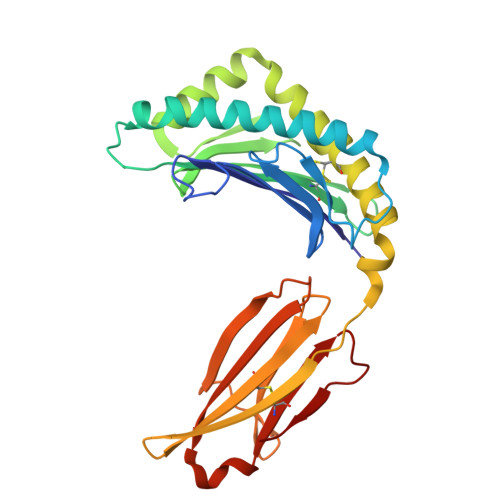
Chicken YF1.7*1 presenting myristoylated peptide derived from tegument protein MDV023


Experimental Data Snapshot

  • Method: X-RAY DIFFRACTION
  • Resolution: 2.08 Å
  • R-Value Free: 
    0.243 (Depositor), 0.243 (DCC) 
  • R-Value Work: 
    0.186 (Depositor), 0.195 (DCC) 
  • R-Value Observed: 
    0.189 (Depositor) 

Starting Model: experimental
View more details

wwPDB Validation   3D Report Full Report


Ligand Structure Quality Assessment 


This is version 1.0 of the entry. See complete history


Literature

Molecular basis for presentation of N-myristoylated peptides by the chicken YF1∗7.1 molecule.

Khandokar, Y.Cheng, T.Y.Wang, C.J.H.Cao, T.P.Nagampalli, R.S.K.Sivaraman, K.K.Van Rhijn, I.Rossjohn, J.Moody, D.B.Nours, J.L.

(2025) J Biological Chem 301: 110253-110253

  • DOI: https://doi.org/10.1016/j.jbc.2025.110253
  • Primary Citation of Related Structures:  
    9EB2, 9EB3, 9EB4, 9EB5, 9EB6

  • PubMed Abstract: 

    Major histocompatibility complex I (MHC-I) and MHC-I-like molecules play a central role in mediating immunity. Through their conservation across all taxa of jawed vertebrates, the MHC-I-like proteins have adapted to present non-peptidic antigens to distinct T cell populations. While our understanding of the structure-function relationship of MHC-I and MHC-I-like molecules in humans and mice is well established, the nature of the antigens presented by MHC-I- like molecules in "non-model" species remains unclear. Here, using a mammalian recombinant expression system combined with mass spectrometry approaches, we identified N-myristoylated peptides as endogenous ligands for the chicken MHC-I-like protein YF1∗7.1. Given the importance of N-myristoylation in viral pathogenesis, we determined the crystal structure of YF1∗7.1 in complex with two N-myristoylated peptides derived from Marek's disease virus (MDV), demonstrating the molecular basis that underpins the presentation of N-myristoylated peptides from MDV, a highly contagious and fatal viral neoplastic disease in chickens. Thus, the identified ligands are distinct from unmodified peptides found in classical MHC-I and -II as well as diverse amphipathic lipids captured by CD1 proteins. Collectively, our study lays the foundation for further molecular and functional characterization of YF1∗7.1 and more broadly of the role of the MHC-I encoded by the MHC-Y gene cluster in protection against highly contagious viral neoplastic diseases in chickens.


  • Organizational Affiliation

    Infection and Immunity Program and Department of Biochemistry and Molecular Biology, Biomedicine Discovery Institute, Monash University, Clayton, Victoria, Australia.


Macromolecules
Find similar proteins by:  (by identity cutoff)  |  3D Structure
Entity ID: 1
MoleculeChains Sequence LengthOrganismDetailsImage
MHC Rfp-Y class I alpha chain270Gallus gallusMutation(s): 0 
Gene Names: YFV
UniProt
Find proteins for Q9BCW3 (Gallus gallus)
Explore Q9BCW3 
Go to UniProtKB:  Q9BCW3
Entity Groups  
Sequence Clusters30% Identity50% Identity70% Identity90% Identity95% Identity100% Identity
UniProt GroupQ9BCW3
Sequence Annotations
Expand
  • Reference Sequence
Find similar proteins by:  (by identity cutoff)  |  3D Structure
Entity ID: 2
MoleculeChains Sequence LengthOrganismDetailsImage
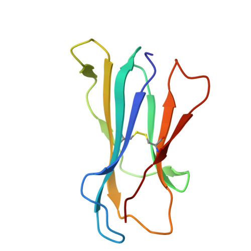
Beta-2-microglobulin98Gallus gallusMutation(s): 0 
Gene Names: B2M
UniProt
Find proteins for P21611 (Gallus gallus)
Explore P21611 
Go to UniProtKB:  P21611
Entity Groups  
Sequence Clusters30% Identity50% Identity70% Identity90% Identity95% Identity100% Identity
UniProt GroupP21611
Sequence Annotations
Expand
  • Reference Sequence

Find similar proteins by:  Sequence   |   3D Structure  

Entity ID: 3
MoleculeChains Sequence LengthOrganismDetailsImage
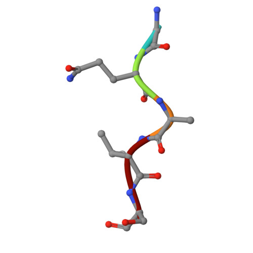
5mer derived from a tegument protein in MDVC [auth D]5Marek's disease herpesvirus type 1 strain MD5Mutation(s): 0 
Sequence Annotations
Expand
  • Reference Sequence
Small Molecules
Ligands 5 Unique
IDChains Name / Formula / InChI Key2D Diagram3D Interactions
MYR (Subject of Investigation/LOI)
Query on MYR

Download Ideal Coordinates CCD File 
I [auth D]MYRISTIC ACID
C14 H28 O2
TUNFSRHWOTWDNC-UHFFFAOYSA-N
PEG
Query on PEG

Download Ideal Coordinates CCD File 
D [auth A]DI(HYDROXYETHYL)ETHER
C4 H10 O3
MTHSVFCYNBDYFN-UHFFFAOYSA-N
CL
Query on CL

Download Ideal Coordinates CCD File 
F [auth A],
G [auth A]
CHLORIDE ION
Cl
VEXZGXHMUGYJMC-UHFFFAOYSA-M
MG
Query on MG

Download Ideal Coordinates CCD File 
H [auth B]MAGNESIUM ION
Mg
JLVVSXFLKOJNIY-UHFFFAOYSA-N
NA
Query on NA

Download Ideal Coordinates CCD File 
E [auth A]SODIUM ION
Na
FKNQFGJONOIPTF-UHFFFAOYSA-N
Experimental Data & Validation

Experimental Data

  • Method: X-RAY DIFFRACTION
  • Resolution: 2.08 Å
  • R-Value Free:  0.243 (Depositor), 0.243 (DCC) 
  • R-Value Work:  0.186 (Depositor), 0.195 (DCC) 
  • R-Value Observed: 0.189 (Depositor) 
Space Group: I 1 2 1
Unit Cell:
Length ( Å )Angle ( ˚ )
a = 57.116α = 90
b = 55.429β = 101.43
c = 133.412γ = 90
Software Package:
Software NamePurpose
MOSFLMdata reduction
Aimlessdata scaling
PHASERphasing
REFMACrefinement
PHENIXrefinement

Structure Validation

View Full Validation Report


Ligand Structure Quality Assessment 


Entry History & Funding Information

Deposition Data


Funding OrganizationLocationGrant Number
National Health and Medical Research Council (NHMRC, Australia)Australia2008616
Monash University/ARC Centre of Excellence in Advanced Molecular Imaging AllianceAustraliaCE140100011

Revision History  (Full details and data files)

  • Version 1.0: 2025-07-23
    Type: Initial release