9EC2 | pdb_00009ec2

Crystal structure of SAMHD1 dimer bound to an inhibitor obtained from high-throughput chemical tethering to the guanine antiviral acyclovir


Experimental Data Snapshot

  • Method: X-RAY DIFFRACTION
  • Resolution: 2.72 Å
  • R-Value Free: 
    0.234 (Depositor), 0.232 (DCC) 
  • R-Value Work: 
    0.188 (Depositor), 0.188 (DCC) 
  • R-Value Observed: 
    0.190 (Depositor) 

Starting Model: experimental
View more details

wwPDB Validation   3D Report Full Report


Ligand Structure Quality Assessment 


This is version 1.1 of the entry. See complete history


Literature

Inhibitors of SAMHD1 Obtained from Chemical Tethering to the Guanine Antiviral Acyclovir.

Egleston, M.Bhat, S.Howlader, A.H.Bianchet, M.A.Liu, Y.Lopez Rovira, L.M.Smith, B.Greenberg, M.M.Stivers, J.T.

(2025) Biochemistry 64: 1109-1120

  • DOI: https://doi.org/10.1021/acs.biochem.4c00854
  • Primary Citation of Related Structures:  
    9EC2

  • PubMed Abstract: 

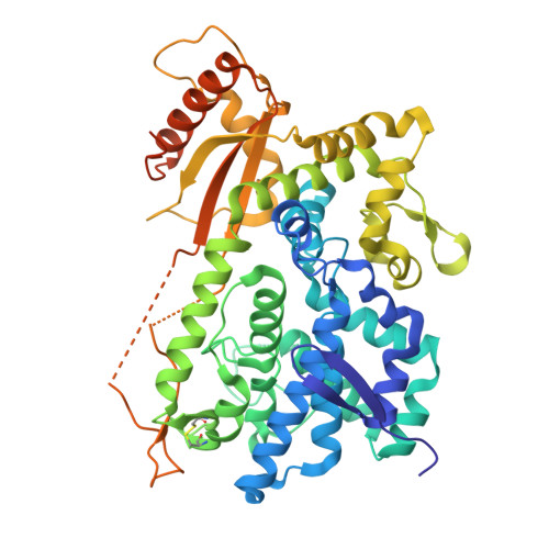
    Sterile alpha motif histidine-aspartate domain protein 1 (SAMHD1) is an enzyme with diverse activities. Its dNTPase activity degrades all canonical dNTPs and many anticancer nucleoside drugs, while its single-stranded nucleic acid binding activity promotes DNA repair and RNA homeostasis in cells. These functions require guanine nucleotide binding to a specific allosteric site (A1) on the enzyme. We previously described how the activities of SAMHD1 could be inhibited in vitro with fragment-based inhibitor design, using dGMP as a targeting fragment for the A1 site. However, these dGMP-tethered inhibitors had poor cell permeability due to the charged guanine monophosphate group. Here, we describe a new approach where the amino form of the guanine acyclic nucleoside acyclovir (NH 2 -ACV) is used as the targeting fragment, allowing facile coupling to activated carboxylic acids (R-COOH), either directly or using linkers. This approach generates a neutral amide instead of charged monophosphate attachment points. High-throughput screening of a ∼375 compound carboxylic acid library identified two compounds ( 8 , 11 ) with similar micromolar affinities for SAMHD1. Compound 11 was obtained by direct coupling to NH 2 -ACV, while compound 8 used a five-carbon linker. Both inhibitors had the same dibromonaphthol component from the carboxylic acid library screen. A crystal structure of a complex between SAMHD1 and 8, combined with computational models of bound 11, suggest how the dibromonaphthol promotes binding. The findings establish that guanine-based inhibitors targeting the A1 site do not require nucleotide or cyclic nucleoside structural elements. This guanine site targeting strategy is highly amenable to further chemical optimization.


  • Organizational Affiliation
    • Department of Pharmacology and Molecular Sciences, Johns Hopkins University School of Medicine, 725 North Wolfe Street, Baltimore, Maryland 21205, United States.

Macromolecules
Find similar proteins by:  (by identity cutoff)  |  3D Structure
Entity ID: 1
MoleculeChains Sequence LengthOrganismDetailsImage
Deoxynucleoside triphosphate triphosphohydrolase SAMHD1
A, B, C, D
516Homo sapiensMutation(s): 0 
Gene Names: SAMHD1MOP5
EC: 3.1.5
UniProt & NIH Common Fund Data Resources
Find proteins for Q9Y3Z3 (Homo sapiens)
Explore Q9Y3Z3 
Go to UniProtKB:  Q9Y3Z3
PHAROS:  Q9Y3Z3
GTEx:  ENSG00000101347 
Entity Groups  
Sequence Clusters30% Identity50% Identity70% Identity90% Identity95% Identity100% Identity
UniProt GroupQ9Y3Z3
Sequence Annotations
Expand
  • Reference Sequence
Small Molecules
Ligands 2 Unique
IDChains Name / Formula / InChI Key2D Diagram3D Interactions
A1BHL
Query on A1BHL

Download Ideal Coordinates CCD File 
E [auth A],
G [auth B],
H [auth B],
J [auth C]
N-[5-({2-[(2-amino-6-oxo-1,6-dihydro-9H-purin-9-yl)methoxy]ethyl}amino)-5-oxopentyl]-4,7-dibromo-3-hydroxynaphthalene-2-carboxamide
C24 H25 Br2 N7 O5
OFCSNXRJBLPABY-UHFFFAOYSA-N
FE
Query on FE

Download Ideal Coordinates CCD File 
F [auth A],
I [auth B],
K [auth C],
L [auth D]
FE (III) ION
Fe
VTLYFUHAOXGGBS-UHFFFAOYSA-N
Experimental Data & Validation

Experimental Data

  • Method: X-RAY DIFFRACTION
  • Resolution: 2.72 Å
  • R-Value Free:  0.234 (Depositor), 0.232 (DCC) 
  • R-Value Work:  0.188 (Depositor), 0.188 (DCC) 
  • R-Value Observed: 0.190 (Depositor) 
Space Group: P 1
Unit Cell:
Length ( Å )Angle ( ˚ )
a = 81.347α = 73.047
b = 94.274β = 71.481
c = 96.579γ = 65.203
Software Package:
Software NamePurpose
PHENIXrefinement
autoPROCdata reduction
Aimlessdata scaling
MOLREPphasing

Structure Validation

View Full Validation Report


Ligand Structure Quality Assessment 


Entry History & Funding Information

Deposition Data


Funding OrganizationLocationGrant Number
National Institutes of Health/National Institute of General Medical Sciences (NIH/NIGMS)United StatesGM056834
National Institutes of Health/National Cancer Institute (NIH/NCI)United StatesCA233567
National Institutes of Health/National Institute of General Medical Sciences (NIH/NIGMS)United StatesGM131736
National Institutes of Health/National Institute of General Medical Sciences (NIH/NIGMS)United StatesGM114259

Revision History  (Full details and data files)

  • Version 1.0: 2025-03-12
    Type: Initial release
  • Version 1.1: 2025-03-19
    Changes: Database references