9ER4 | pdb_00009er4

Room temperature structure of Glycine max phyA in Pr


Experimental Data Snapshot

  • Method: X-RAY DIFFRACTION
  • Resolution: 2.20 Å
  • R-Value Free: 
    0.241 (Depositor), 0.240 (DCC) 
  • R-Value Work: 
    0.204 (Depositor), 0.204 (DCC) 
  • R-Value Observed: 
    0.206 (Depositor) 

Starting Model: experimental
View more details

wwPDB Validation   3D Report Full Report


Ligand Structure Quality Assessment 


This is version 1.2 of the entry. See complete history


Literature

Pr and Pfr structures of plant phytochrome A.

Nagano, S.von Stetten, D.Guan, K.Chen, P.Y.Song, C.Barends, T.Weiss, M.S.Feiler, C.G.Dorner, K.de Diego Martinez, I.Schubert, R.Bielecki, J.Brings, L.Han, H.Kharitonov, K.Kim, C.Kloos, M.Koliyadu, J.C.P.Koua, F.H.M.Round, E.Sarma, A.Sato, T.Schmidt, C.Valerio, J.Wrona, A.Schulz, J.de Wijn, R.Letrun, R.Bean, R.Mancuso, A.Heyne, K.Hughes, J.

(2025) Nat Commun 16: 5319-5319

  • DOI: https://doi.org/10.1038/s41467-025-60738-w
  • Primary Citation of Related Structures:  
    8R44, 8R45, 9ER4, 9F4I, 9QZT

  • PubMed Abstract: 

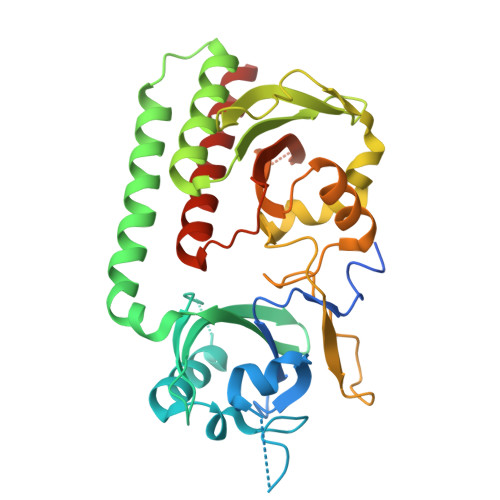
    Phytochromes are biliprotein photoreceptors widespread amongst microorganisms and ubiquitous in plants where they control developmental processes as diverse as germination, stem elongation and floral induction through the photoconversion of inactive Pr to the Pfr signalling state. Here we report crystal structures of the chromophore-binding module of soybean phytochrome A, including ~2.2 Å XFEL structures of Pr and Pfr at ambient temperature and high resolution cryogenic structures of Pr. In the Pfr structure, the chromophore is exposed to the medium, the D-ring remaining α-facial following the likely clockwise photoflip. The chromophore shifts within its pocket, while its propionate side chains, their partners as well as three neighbouring tyrosines shift radically. Helices near the chromophore show substantial shifts that might represent components of the light signal. These changes reflect those in bacteriophytochromes despite their quite different signalling mechanisms, implying that fundamental aspects of phytochrome photoactivation have been repurposed for photoregulation in the eukaryotic plant.


  • Organizational Affiliation

    Institute for Plant Physiology, Justus Liebig University, Giessen, Germany.


Macromolecules
Find similar proteins by:  (by identity cutoff)  |  3D Structure
Entity ID: 1
MoleculeChains Sequence LengthOrganismDetailsImage
Phytochrome A-2
A, B
359Glycine maxMutation(s): 0 
Gene Names: PHYA2GLYMA_20G090000
UniProt
Find proteins for B4YB07 (Glycine max)
Explore B4YB07 
Go to UniProtKB:  B4YB07
Entity Groups  
Sequence Clusters30% Identity50% Identity70% Identity90% Identity95% Identity100% Identity
UniProt GroupB4YB07
Sequence Annotations
Expand
  • Reference Sequence
Experimental Data & Validation

Experimental Data

  • Method: X-RAY DIFFRACTION
  • Resolution: 2.20 Å
  • R-Value Free:  0.241 (Depositor), 0.240 (DCC) 
  • R-Value Work:  0.204 (Depositor), 0.204 (DCC) 
  • R-Value Observed: 0.206 (Depositor) 
Space Group: P 1 21 1
Unit Cell:
Length ( Å )Angle ( ˚ )
a = 56.45α = 90
b = 115.05β = 92.68
c = 69.8γ = 90
Software Package:
Software NamePurpose
PHENIXrefinement
CrystFELdata reduction
CrystFELdata scaling
PHASERphasing

Structure Validation

View Full Validation Report


Ligand Structure Quality Assessment 


Entry History & Funding Information

Deposition Data

  • Released Date: 2025-01-08 
  • Deposition Author(s): Nagano, S.

Funding OrganizationLocationGrant Number
German Research Foundation (DFG)GermanySFB1078

Revision History  (Full details and data files)

  • Version 1.0: 2025-01-08
    Type: Initial release
  • Version 1.1: 2025-02-19
    Changes: Database references
  • Version 1.2: 2025-07-02
    Changes: Database references