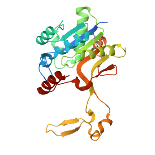
9FTC | pdb_00009ftc

Aeromonas caviae CMP-Pse5Ac7Ac synthetase in the presence of CMP-Pse5Ac7Ac.


Experimental Data Snapshot

  • Method: X-RAY DIFFRACTION
  • Resolution: 1.50 Å
  • R-Value Free: 
    0.185 (Depositor), 0.185 (DCC) 
  • R-Value Work: 
    0.167 (Depositor), 0.167 (DCC) 

Starting Model: experimental
View more details

wwPDB Validation   3D Report Full Report


Ligand Structure Quality Assessment 


This is version 1.2 of the entry. See complete history


Literature

Structural dissection of the CMP-pseudaminic acid synthetase, PseF.

Keenan, T.Cowan, A.R.Flack, E.K.P.Hatton, N.E.Walklett, A.J.Thomas, G.H.Hemsworth, G.R.Fascione, M.A.

(2024) Structure 32: 2399

  • DOI: https://doi.org/10.1016/j.str.2024.09.017
  • Primary Citation of Related Structures:  
    9FTB, 9FTC

  • PubMed Abstract: 

    Pseudaminic acid is a non-mammalian sugar found in the surface glycoconjugates of many bacteria, including several human pathogens, and is a virulence factor thought to facilitate immune evasion. The final step in the biosynthesis of the nucleotide activated form of the sugar, CMP-Pse5Ac7Ac is performed by a CMP-Pse5Ac7Ac synthetase (PseF). Here we present the biochemical and structural characterization of PseF from Aeromonas caviae (AcPseF), with AcPseF displaying metal-dependent activity over a broad pH and temperature range. Upon binding to CMP-Pse5Ac7Ac, AcPseF undergoes dynamic movements akin to other CMP-ulosonic acid synthetases. The enzyme clearly discriminates Pse5Ac7Ac from other ulosonic acids, through active site interactions with side-chain functional groups and by positioning the molecule in a hydrophobic pocket. Finally, we show that AcPseF binds the CMP-Pse5Ac7Ac side chain in the lowest energy conformation, a trend that we observed in the structures of other enzymes of this class.


  • Organizational Affiliation
    • Department of Chemistry, University of York, York YO10 5DD, UK.

Macromolecules
Find similar proteins by:  (by identity cutoff)  |  3D Structure
Entity ID: 1
MoleculeChains Sequence LengthOrganismDetailsImage
NeuA
A, B
248Aeromonas caviaeMutation(s): 0 
Gene Names: neuA
UniProt
Find proteins for Q9R9S4 (Aeromonas caviae)
Explore Q9R9S4 
Go to UniProtKB:  Q9R9S4
Entity Groups  
Sequence Clusters30% Identity50% Identity70% Identity90% Identity95% Identity100% Identity
UniProt GroupQ9R9S4
Sequence Annotations
Expand
  • Reference Sequence
Small Molecules
Ligands 1 Unique
IDChains Name / Formula / InChI Key2D Diagram3D Interactions
A1IF6 (Subject of Investigation/LOI)
Query on A1IF6

Download Ideal Coordinates CCD File 
C [auth A],
D [auth B]
(2~{R},4~{S},5~{S},6~{S})-5-acetamido-6-[(1~{S},2~{S})-1-acetamido-2-oxidanyl-propyl]-2-[[(2~{R},3~{S},4~{R},5~{R})-5-(4-azanyl-2-oxidanylidene-pyrimidin-1-yl)-3,4-bis(oxidanyl)oxolan-2-yl]methoxy-oxidanyl-phosphoryl]oxy-4-oxidanyl-oxane-2-carboxylic acid
C22 H34 N5 O15 P
XTZJKGIMUFZFBV-STPXTTIVSA-N
Experimental Data & Validation

Experimental Data

  • Method: X-RAY DIFFRACTION
  • Resolution: 1.50 Å
  • R-Value Free:  0.185 (Depositor), 0.185 (DCC) 
  • R-Value Work:  0.167 (Depositor), 0.167 (DCC) 
Space Group: P 1 21 1
Unit Cell:
Length ( Å )Angle ( ˚ )
a = 42.574α = 90
b = 79.007β = 92.46
c = 74.039γ = 90
Software Package:
Software NamePurpose
REFMACrefinement
Aimlessdata scaling
PHASERphasing
XDSdata reduction

Structure Validation

View Full Validation Report


Ligand Structure Quality Assessment 


Entry History & Funding Information

Deposition Data


Funding OrganizationLocationGrant Number
European Research Council (ERC)European Union101044024

Revision History  (Full details and data files)

  • Version 1.0: 2024-10-09
    Type: Initial release
  • Version 1.1: 2024-10-30
    Changes: Database references
  • Version 1.2: 2024-12-18
    Changes: Database references