


Crystal structure of marine sulfatase (OpSulf1) from Ochrovirga pacifica
Solanki, V.A., Krull, J., Hehemann, J.H.To be published.
Experimental Data Snapshot
Starting Model: in silico
View more details
wwPDB Validation 3D Report Full Report

Entity ID: 1 | |||||
|---|---|---|---|---|---|
| Molecule | Chains | Sequence Length | Organism | Details | Image |
| Sulfatase | 519 | Ochrovirga pacifica | Mutation(s): 0 Gene Names: Sulfatase | 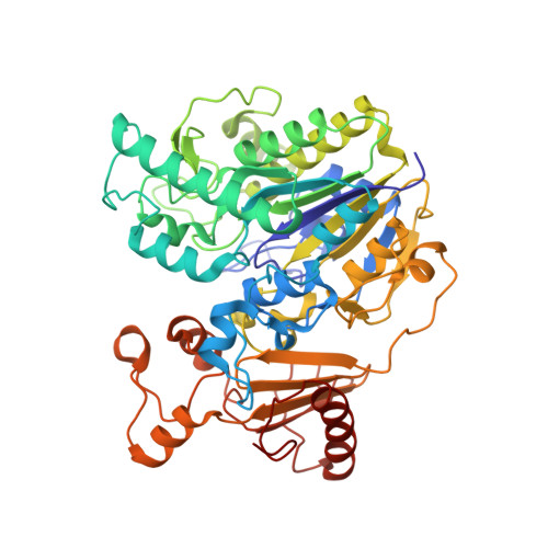 | |
UniProt | |||||
Find proteins for A0AAX7FM35 (Ochrovirga pacifica) Explore A0AAX7FM35 Go to UniProtKB: A0AAX7FM35 | |||||
Entity Groups | |||||
| Sequence Clusters | 30% Identity50% Identity70% Identity90% Identity95% Identity100% Identity | ||||
| UniProt Group | A0AAX7FM35 | ||||
Sequence AnnotationsExpand | |||||
| |||||
| Ligands 1 Unique | |||||
|---|---|---|---|---|---|
| ID | Chains | Name / Formula / InChI Key | 2D Diagram | 3D Interactions | |
| MG (Subject of Investigation/LOI) Query on MG | E [auth A], F [auth B], G [auth C], H [auth D] | MAGNESIUM ION Mg JLVVSXFLKOJNIY-UHFFFAOYSA-N |  | ||
| Length ( Å ) | Angle ( ˚ ) |
|---|---|
| a = 115.268 | α = 90 |
| b = 115.268 | β = 90 |
| c = 169.831 | γ = 90 |
| Software Name | Purpose |
|---|---|
| REFMAC | refinement |
| Aimless | data scaling |
| PHASER | phasing |
| XDS | data reduction |
| Funding Organization | Location | Grant Number |
|---|---|---|
| German Research Foundation (DFG) | Germany | HE 7217/2-3 |


