9GA6 | pdb_00009ga6

The crystal structure of human Annexin A4 derived from crystals grown in 40 mM of CaCl2


Experimental Data Snapshot

  • Method: X-RAY DIFFRACTION
  • Resolution: 1.27 Å
  • R-Value Free: 
    0.189 (Depositor), 0.195 (DCC) 
  • R-Value Work: 
    0.174 (Depositor), 0.183 (DCC) 

Starting Model: experimental
View more details

wwPDB Validation   3D Report Full Report


Ligand Structure Quality Assessment 


This is version 1.0 of the entry. See complete history


Literature

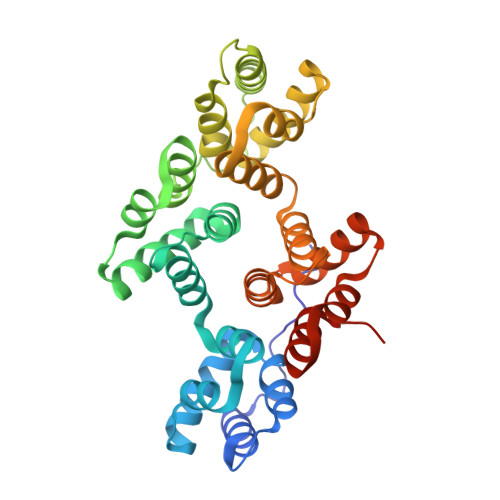
Computational, crystallographic, and biophysical characterizations provide insights into calcium and phosphate binding by human annexin A4.

Di Micco, S.Scala, M.C.Sala, M.Barra, G.Ghilardi, O.Campiglia, P.Bifulco, G.Vitagliano, L.Ruggiero, A.

(2025) Int J Biol Macromol 308: 142600-142600

  • DOI: https://doi.org/10.1016/j.ijbiomac.2025.142600
  • Primary Citation of Related Structures:  
    9GA6, 9GA7, 9GA8

  • PubMed Abstract: 

    The members of the annexin family are proteins involved in important biological processes that share a common propensity, mediated by the binding of calcium, to interact with membranes. Despite the remarkable amount of literature reports on these proteins several aspects of their functionality remain obscure. Considering the importance of the pH in modulating annexin activities, we here reassessed the pH dependency (range 4.6-7.4) of the binding of the calcium by human annexin A4 (hAnxA4) and determined its structure from crystals obtained in acidic conditions at nearly atomic resolution in media containing different calcium concentrations. The interactions of calcium ions with hAnxA4 were studied using isothermal titration calorimetry measurements and molecular dynamics simulations. Present solution data corroborate and quantify the pH dependence of the binding of calcium to hAnxA4. Moreover, crystallographic structures provide a clear ranking of the metal affinity of the hAnxA4 calcium binding sites. These findings have been extended by performing computational studies that provide information on the binding affinity of the different calcium sites that are in good agreement with the crystallographic data. Crystallographic data highlight the occurrence of unexpected clusterings of positively charged arginine residues that can cooperate for the binding of the phospholipid phosphate moieties. These crystallographic data integrated with molecular dynamics simulations provide an atomic-level description of the local conformational changes associated with calcium release and upload. Interestingly, docking analyses demonstrate the optimal juxtaposition of these arginine residues and calcium ions to correctly anchor phosphatidylserine.


  • Organizational Affiliation

    European Biomedical Research Institute (EBRIS), Via S. De Renzi 50, 84125 Salerno, Italy.


Macromolecules
Find similar proteins by:  (by identity cutoff)  |  3D Structure
Entity ID: 1
MoleculeChains Sequence LengthOrganismDetailsImage
Annexin A4325Homo sapiensMutation(s): 0 
Gene Names: ANXA4ANX4
UniProt & NIH Common Fund Data Resources
Find proteins for P09525 (Homo sapiens)
Explore P09525 
Go to UniProtKB:  P09525
PHAROS:  P09525
GTEx:  ENSG00000196975 
Entity Groups  
Sequence Clusters30% Identity50% Identity70% Identity90% Identity95% Identity100% Identity
UniProt GroupP09525
Sequence Annotations
Expand
  • Reference Sequence
Small Molecules
Ligands 3 Unique
IDChains Name / Formula / InChI Key2D Diagram3D Interactions
SO4 (Subject of Investigation/LOI)
Query on SO4

Download Ideal Coordinates CCD File 
H [auth A]
I [auth A]
J [auth A]
K [auth A]
L [auth A]
H [auth A],
I [auth A],
J [auth A],
K [auth A],
L [auth A],
M [auth A]
SULFATE ION
O4 S
QAOWNCQODCNURD-UHFFFAOYSA-L
GOL (Subject of Investigation/LOI)
Query on GOL

Download Ideal Coordinates CCD File 
E [auth A],
F [auth A],
G [auth A]
GLYCEROL
C3 H8 O3
PEDCQBHIVMGVHV-UHFFFAOYSA-N
CA (Subject of Investigation/LOI)
Query on CA

Download Ideal Coordinates CCD File 
B [auth A],
C [auth A],
D [auth A]
CALCIUM ION
Ca
BHPQYMZQTOCNFJ-UHFFFAOYSA-N
Experimental Data & Validation

Experimental Data

  • Method: X-RAY DIFFRACTION
  • Resolution: 1.27 Å
  • R-Value Free:  0.189 (Depositor), 0.195 (DCC) 
  • R-Value Work:  0.174 (Depositor), 0.183 (DCC) 
Space Group: P 1 21 1
Unit Cell:
Length ( Å )Angle ( ˚ )
a = 58.742α = 90
b = 39.906β = 93.91
c = 73.456γ = 90
Software Package:
Software NamePurpose
REFMACrefinement
XDSdata reduction
Aimlessdata scaling
PHASERphasing

Structure Validation

View Full Validation Report


Ligand Structure Quality Assessment 


Entry History & Funding Information

Deposition Data


Funding OrganizationLocationGrant Number
Not funded--

Revision History  (Full details and data files)

  • Version 1.0: 2025-06-04
    Type: Initial release