9GFY | pdb_00009gfy

Endothiapepsin in complex with pepstatin soaked at pH 7.6


Experimental Data Snapshot

  • Method: X-RAY DIFFRACTION
  • Resolution: 1.23 Å
  • R-Value Free: 
    0.174 (Depositor), 0.174 (DCC) 
  • R-Value Work: 
    0.148 (Depositor), 0.148 (DCC) 
  • R-Value Observed: 
    0.149 (Depositor) 

Starting Model: experimental
View more details

wwPDB Validation   3D Report Full Report


This is version 1.0 of the entry. See complete history


Literature

Protonation Effects in Protein-Ligand Complexes - A Case Study of Endothiapepsin and Pepstatin A with Computational and Experimental Methods.

Vatheuer, H.Palomino-Hernandez, O.Muller, J.Galonska, P.Glinca, S.Czodrowski, P.

(2025) ChemMedChem 20: e202400953-e202400953

  • DOI: https://doi.org/10.1002/cmdc.202400953
  • Primary Citation of Related Structures:  
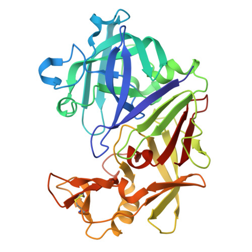
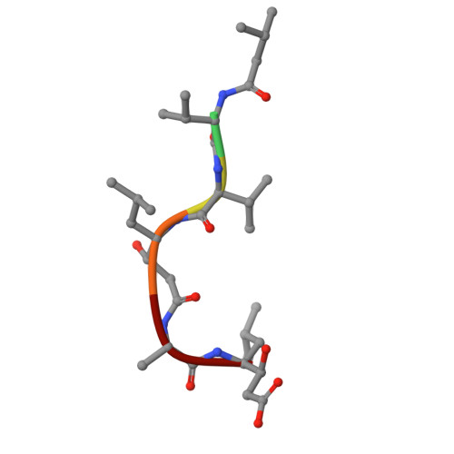
    9GFY

  • PubMed Abstract: 

    Protonation states serve as an essential molecular recognition motif for biological processes. Their correct consideration is key to successful drug design campaigns, since chemoinformatic tools usually deal with default protonation states of ligands and proteins and miss atypical protonation states. The protonation pattern for the Endothiapepsin/PepstatinA (EP/pepA) complex is investigated using different dry lab and wet lab techniques. ITC experiments revealed an uptake of more than one mole of protons upon pepA binding to EP. Since these experiments were performed at physiological conditions (and not at pH=4.6 at which a large variety of crystal structures is available), a novel crystal structure at pH=7.6 was determined. This crystal structure showed that only modest structural changes occur upon increasing the pH value. This lead to computational studies Poisson-Boltzmann calculations and constant pH MD simulation to reveal the exact location of the protonation event. Both computational studies could reveal a significant pKa shift resulting in non-default protonation state and that the catalytic dyad is responsible for the uptake of protons. This study shows that assessing protonation states for two separate systems (protein and ligand) might result in the incorrect assignment of protonation states and hence incorrect calculation of binding energy.


  • Organizational Affiliation

    Chemistry Department, Johannes Gutenberg University, Duesbergweg 10-14, 55128, Mainz, Germany.


Macromolecules
Find similar proteins by:  (by identity cutoff)  |  3D Structure
Entity ID: 1
MoleculeChains Sequence LengthOrganismDetailsImage
Endothiapepsin330Cryphonectria parasiticaMutation(s): 0 
EC: 3.4.23.22
UniProt
Find proteins for P11838 (Cryphonectria parasitica)
Explore P11838 
Go to UniProtKB:  P11838
Entity Groups  
Sequence Clusters30% Identity50% Identity70% Identity90% Identity95% Identity100% Identity
UniProt GroupP11838
Sequence Annotations
Expand
  • Reference Sequence

Find similar proteins by:  Sequence   |   3D Structure  

Entity ID: 2
MoleculeChains Sequence LengthOrganismDetailsImage
Pepstatin6Streptomyces argenteolus subsp. toyonakensisMutation(s): 0 
Sequence Annotations
Expand
  • Reference Sequence
Small Molecules
Ligands 2 Unique
IDChains Name / Formula / InChI Key2D Diagram3D Interactions
1PE
Query on 1PE

Download Ideal Coordinates CCD File 
D [auth A]PENTAETHYLENE GLYCOL
C10 H22 O6
JLFNLZLINWHATN-UHFFFAOYSA-N
DMS
Query on DMS

Download Ideal Coordinates CCD File 
C [auth A]
E [auth A]
F [auth A]
G [auth A]
H [auth A]
C [auth A],
E [auth A],
F [auth A],
G [auth A],
H [auth A],
I [auth A],
J [auth A]
DIMETHYL SULFOXIDE
C2 H6 O S
IAZDPXIOMUYVGZ-UHFFFAOYSA-N
Biologically Interesting Molecules (External Reference) 1 Unique
Experimental Data & Validation

Experimental Data

  • Method: X-RAY DIFFRACTION
  • Resolution: 1.23 Å
  • R-Value Free:  0.174 (Depositor), 0.174 (DCC) 
  • R-Value Work:  0.148 (Depositor), 0.148 (DCC) 
  • R-Value Observed: 0.149 (Depositor) 
Space Group: P 1 21 1
Unit Cell:
Length ( Å )Angle ( ˚ )
a = 45.12α = 90
b = 72.623β = 109.22
c = 52.295γ = 90
Software Package:
Software NamePurpose
PHENIXrefinement
XDSdata reduction
XDSdata scaling
PHASERphasing
Cootmodel building

Structure Validation

View Full Validation Report


Entry History & Funding Information

Deposition Data


Funding OrganizationLocationGrant Number
Not funded--

Revision History  (Full details and data files)

  • Version 1.0: 2025-06-11
    Type: Initial release