9GSO | pdb_00009gso

DNA binding domain of J-DNA Binding Protein 3 (JBP3)


Experimental Data Snapshot

  • Method: X-RAY DIFFRACTION
  • Resolution: 1.61 Å
  • R-Value Free: 
    0.222 (Depositor), 0.227 (DCC) 
  • R-Value Work: 
    0.190 (Depositor), 0.200 (DCC) 

Starting Model: in silico
View more details

wwPDB Validation   3D Report Full Report


This is version 1.1 of the entry. See complete history


Literature

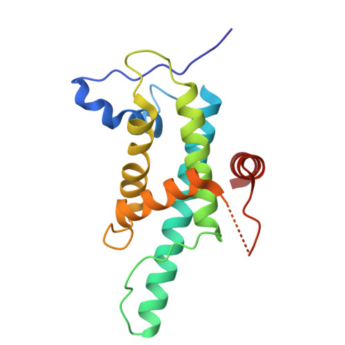
JBP1 and JBP3 have conserved structures but different affinity to base-J.

de Vries, I.Adamopoulos, A.Kazokaite-Adomaitiene, J.Heidebrecht, T.Fish, A.Celie, P.H.N.Joosten, R.P.Perrakis, A.

(2024) J Struct Biol 217: 108161-108161

  • DOI: https://doi.org/10.1016/j.jsb.2024.108161
  • Primary Citation of Related Structures:  
    9GSO, 9GSQ

  • PubMed Abstract: 

    Base-J (β-D-glucopyranosyloxymethyluracil) is an unusual kinetoplastid-specific DNA modification, recognized by base-J containing DNA (J-DNA) binding proteins JBP1 and JBP3. Recognition of J-DNA by both JBP1 and JBP3 takes place by a conserved J-DNA binding domain (JDBD). Here we show that JDBD-JBP3 has about 1,000-fold weaker affinity to base-J than JDBD-JBP1 and discriminates between J-DNA and unmodified DNA with a factor ∼5, whereas JDBD-JBP1 discriminates with a factor ∼10,000. Comparison of the crystal structures of JDBD-JBP3 we present here, with that of the previously characterized JDBD-JBP1, shows a flexible α5-helix that lacks a positively charged patch in JBP3. Mutations removing this positive charge in JDBD-JBP1, resulted in decreased binding affinity relative to wild-type JDBD-JBP1, indicating this patch is involved in DNA binding. We suggest that the α5-helix might rearrange upon JBP1 binding to J-DNA stabilizing the complex. This work contributes to our understanding of how JBPs bind to this unique DNA modification, which may contribute to identifying potential drug targets to end the base-J dependent parasite life cycle.


  • Organizational Affiliation
    • Oncode Institute and Division of Biochemistry at the Netherlands Cancer Institute - Plesmanlaan 121, 1066 CX Amsterdam, the Netherlands.

Macromolecules
Find similar proteins by:  (by identity cutoff)  |  3D Structure
Entity ID: 1
MoleculeChains Sequence LengthOrganismDetailsImage
DNA binding domain of J-DNA Binding Protein 3 (JBP3)177Leishmania tarentolaeMutation(s): 0 
Gene Names: LtaPh_3603800
UniProt
Find proteins for A0A640KTL4 (Leishmania tarentolae)
Explore A0A640KTL4 
Go to UniProtKB:  A0A640KTL4
Entity Groups  
Sequence Clusters30% Identity50% Identity70% Identity90% Identity95% Identity100% Identity
UniProt GroupA0A640KTL4
Sequence Annotations
Expand
  • Reference Sequence
Experimental Data & Validation

Experimental Data

  • Method: X-RAY DIFFRACTION
  • Resolution: 1.61 Å
  • R-Value Free:  0.222 (Depositor), 0.227 (DCC) 
  • R-Value Work:  0.190 (Depositor), 0.200 (DCC) 
Space Group: P 2 21 21
Unit Cell:
Length ( Å )Angle ( ˚ )
a = 46.828α = 90
b = 59.678β = 90
c = 67.288γ = 90
Software Package:
Software NamePurpose
REFMACrefinement
autoPROCdata reduction
Aimlessdata scaling
MOLREPphasing
PDB-REDOrefinement

Structure Validation

View Full Validation Report


Entry History & Funding Information

Deposition Data


Funding OrganizationLocationGrant Number
Netherlands Organisation for Scientific Research (NWO)Netherlands714.014.002

Revision History  (Full details and data files)

  • Version 1.0: 2024-09-25
    Type: Initial release
  • Version 1.1: 2025-01-22
    Changes: Database references, Structure summary