9GV7 | pdb_00009gv7

Structure of reverse docking TCR in complex with peptide-HLA


Experimental Data Snapshot

  • Method: X-RAY DIFFRACTION
  • Resolution: 1.86 Å
  • R-Value Free: 
    0.212 (Depositor), 0.221 (DCC) 
  • R-Value Work: 
    0.180 (Depositor), 0.193 (DCC) 
  • R-Value Observed: 
    0.182 (Depositor) 

Starting Models: experimental
View more details

wwPDB Validation   3D Report Full Report


This is version 1.1 of the entry. See complete history


Literature

Determining T-cell receptor binding orientation and Peptide-HLA interactions using cross-linking mass spectrometry.

Powell, T.Karuppiah, V.Shaikh, S.A.Pengelly, R.Mai, N.Barnbrook, K.Sharma, A.Harper, S.Ebner, M.Creese, A.J.

(2025) J Biological Chem 301: 108445-108445

  • DOI: https://doi.org/10.1016/j.jbc.2025.108445
  • Primary Citation of Related Structures:  
    9GV6, 9GV7

  • PubMed Abstract: 

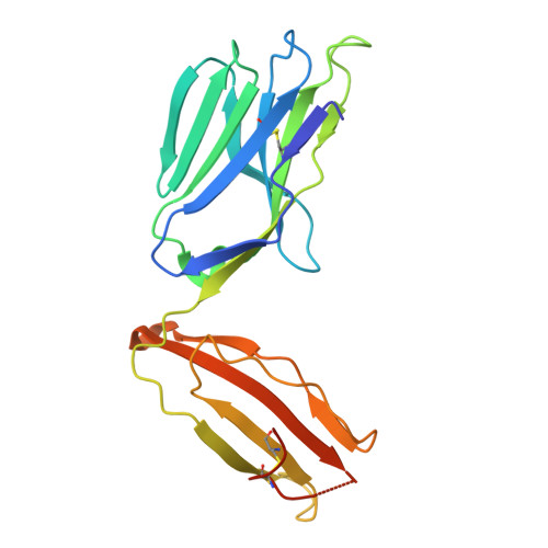
    T cell receptors (TCRs) recognize specific peptides presented by human leukocyte antigens (HLAs) on the surface of antigen-presenting cells and are involved in fighting pathogens and cancer surveillance. Canonical docking orientation of TCRs to their target peptide-HLAs (pHLAs) is essential for T cell activation, with reverse binding TCRs lacking functionality. TCR binding geometry and molecular interaction footprint with pHLAs are typically obtained by determining the crystal structure. Here, we describe the use of a cross-linking tandem mass spectrometry (XL-MS/MS) method to decipher the binding orientation of several TCRs to their target pHLAs. Cross-linking sites were localized to specific residues and their molecular interactions showed differentiation between TCRs binding in canonical or reverse orientations. Structural prediction and crystal structure determination of two TCR-pHLA complexes validated these findings. The XL-MS/MS method described herein offers a faster and simpler approach for elucidating TCR-pHLA binding orientation and interactions.


  • Organizational Affiliation
    • Immunocore Limited, Abingdon, United Kingdom. Electronic address: thomas.powell@immunocore.com.

Macromolecules
Find similar proteins by:  (by identity cutoff)  |  3D Structure
Entity ID: 1
MoleculeChains Sequence LengthOrganismDetailsImage
MHC class I antigen276Homo sapiensMutation(s): 0 
Gene Names: HLA-A
UniProt
Find proteins for A0A5B8RNS7 (Homo sapiens)
Explore A0A5B8RNS7 
Go to UniProtKB:  A0A5B8RNS7
Entity Groups  
Sequence Clusters30% Identity50% Identity70% Identity90% Identity95% Identity100% Identity
UniProt GroupA0A5B8RNS7
Sequence Annotations
Expand
  • Reference Sequence
Find similar proteins by:  (by identity cutoff)  |  3D Structure
Entity ID: 2
MoleculeChains Sequence LengthOrganismDetailsImage
Beta-2-microglobulin100Homo sapiensMutation(s): 0 
Gene Names: B2MCDABP0092HDCMA22P
UniProt & NIH Common Fund Data Resources
Find proteins for P61769 (Homo sapiens)
Explore P61769 
Go to UniProtKB:  P61769
PHAROS:  P61769
GTEx:  ENSG00000166710 
Entity Groups  
Sequence Clusters30% Identity50% Identity70% Identity90% Identity95% Identity100% Identity
UniProt GroupP61769
Sequence Annotations
Expand
  • Reference Sequence

Find similar proteins by:  Sequence   |   3D Structure  

Entity ID: 3
MoleculeChains Sequence LengthOrganismDetailsImage
Peptide11Homo sapiensMutation(s): 0 
Entity Groups  
Sequence Clusters30% Identity50% Identity70% Identity90% Identity95% Identity100% Identity
Sequence Annotations
Expand
  • Reference Sequence
Find similar proteins by:  (by identity cutoff)  |  3D Structure
Entity ID: 4
MoleculeChains Sequence LengthOrganismDetailsImage
TCR Alpha200Homo sapiensMutation(s): 0 
Entity Groups  
Sequence Clusters30% Identity50% Identity70% Identity90% Identity95% Identity100% Identity
Sequence Annotations
Expand
  • Reference Sequence
Find similar proteins by:  (by identity cutoff)  |  3D Structure
Entity ID: 5
MoleculeChains Sequence LengthOrganismDetailsImage
TCR Beta244Homo sapiensMutation(s): 0 
Entity Groups  
Sequence Clusters30% Identity50% Identity70% Identity90% Identity95% Identity100% Identity
Sequence Annotations
Expand
  • Reference Sequence
Experimental Data & Validation

Experimental Data

  • Method: X-RAY DIFFRACTION
  • Resolution: 1.86 Å
  • R-Value Free:  0.212 (Depositor), 0.221 (DCC) 
  • R-Value Work:  0.180 (Depositor), 0.193 (DCC) 
  • R-Value Observed: 0.182 (Depositor) 
Space Group: P 65
Unit Cell:
Length ( Å )Angle ( ˚ )
a = 144.13α = 90
b = 144.13β = 90
c = 106.79γ = 120
Software Package:
Software NamePurpose
REFMACrefinement
xia2.multiplexdata reduction
xia2.multiplexdata scaling
PHASERphasing

Structure Validation

View Full Validation Report


Entry History & Funding Information

Deposition Data


Funding OrganizationLocationGrant Number
Other privateUnited Kingdom--

Revision History  (Full details and data files)

  • Version 1.0: 2025-04-23
    Type: Initial release
  • Version 1.1: 2025-04-30
    Changes: Database references