9HJ2 | pdb_00009hj2

Structure of P167S/D240G/D172A/S104G/H184R BlaC from Mycobacterium tuberculosis


Experimental Data Snapshot

  • Method: X-RAY DIFFRACTION
  • Resolution: 1.60 Å
  • R-Value Free: 
    0.252 (Depositor), 0.256 (DCC) 
  • R-Value Work: 
    0.232 (Depositor), 0.237 (DCC) 
  • R-Value Observed: 
    0.233 (Depositor) 

Starting Model: experimental
View more details

wwPDB Validation   3D Report Full Report


This is version 1.0 of the entry. See complete history


Literature

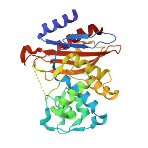
Directed evolution of a beta-lactamase samples a wide variety of conformational states.

Sun, J.Timmer, M.Brunle, S.Boyle, A.L.Ubbink, M.

(2025) Protein Sci 34: e70322-e70322

  • DOI: https://doi.org/10.1002/pro.70322
  • Primary Citation of Related Structures:  
    9HCK, 9HIT, 9HJ2

  • PubMed Abstract: 

    In directed evolution, enzyme activity is improved in successive generations of laboratory evolution, which can be described by a simple stepwise climb toward a peak in the fitness landscape. In a naive model of evolution, it can be assumed that each enzyme variant along this path is in a single, well-defined state that differs slightly from the previous one. We analyzed the structural changes in mutants of the β-lactamase BlaC from Mycobacterium tuberculosis obtained via directed evolution for increased ceftazidime hydrolysis activity. Crystal structures of three successive mutants only show an increase in the dynamics of a loop that lines the active site (Ω-loop), enabling better access of the large substrate. However, NMR spectra of wild type and nine mutants of different branches of the directed evolution experiment show a much more diverse and complex picture of the conformational effects. Many mutants show micro-millisecond dynamics for certain regions and most show peak doubling, indicative of two or more conformations being populated. Thus, the straightforward climb to increased ceftazidime activity in the fitness landscape masks a complex trajectory in the conformational landscape, emphasizing the complex and epistatic interplay that single mutations can have on the structure and dynamics of enzymes.


  • Organizational Affiliation
    • Macromolecular Biochemistry, Leiden Institute of Chemistry, Leiden University, Leiden, The Netherlands.

Macromolecules
Find similar proteins by:  (by identity cutoff)  |  3D Structure
Entity ID: 1
MoleculeChains Sequence LengthOrganismDetailsImage
Beta-lactamaseA,
B [auth C]
265Mycobacterium tuberculosisMutation(s): 3 
Gene Names: blaAblaCA4S10_02181DKC2_2191DSI80_06165ERS007661_01447ERS007681_02835ERS007718_02594ERS007741_00391ERS053720_02349
EC: 3.5.2.6
UniProt
Find proteins for A0A045INV3 (Mycobacterium tuberculosis)
Explore A0A045INV3 
Go to UniProtKB:  A0A045INV3
Entity Groups  
Sequence Clusters30% Identity50% Identity70% Identity90% Identity95% Identity100% Identity
UniProt GroupA0A045INV3
Sequence Annotations
Expand
  • Reference Sequence
Experimental Data & Validation

Experimental Data

  • Method: X-RAY DIFFRACTION
  • Resolution: 1.60 Å
  • R-Value Free:  0.252 (Depositor), 0.256 (DCC) 
  • R-Value Work:  0.232 (Depositor), 0.237 (DCC) 
  • R-Value Observed: 0.233 (Depositor) 
Space Group: P 1 21 1
Unit Cell:
Length ( Å )Angle ( ˚ )
a = 53.827α = 90
b = 57.27β = 95.71
c = 76.927γ = 90
Software Package:
Software NamePurpose
REFMACrefinement
XDSdata reduction
Aimlessdata scaling
MOLREPphasing

Structure Validation

View Full Validation Report


Entry History & Funding Information

Deposition Data


Funding OrganizationLocationGrant Number
Chinese Academy of SciencesChina--

Revision History  (Full details and data files)

  • Version 1.0: 2025-10-22
    Type: Initial release