Enhancing the activity of gamma-hydroxy lactone derivatives as innovative peroxisome proliferator-activated receptor gamma non-agonists inhibiting cyclin-dependent kinase 5-mediated phosphorylation.
Cazzaniga, G., Capelli, D., Montanari, R., Fassi, E.M.A., Grazioso, G., Tresoldi, A., Rinaldi, F., Calleri, E., Bassanini, I., Romeo, S., Garofalo, M., Mori, M., Meneghetti, F., Villa, S.(2025) Eur J Med Chem 292: 117657-117657
- PubMed: 40318479
- DOI: https://doi.org/10.1016/j.ejmech.2025.117657
- Primary Citation of Related Structures:
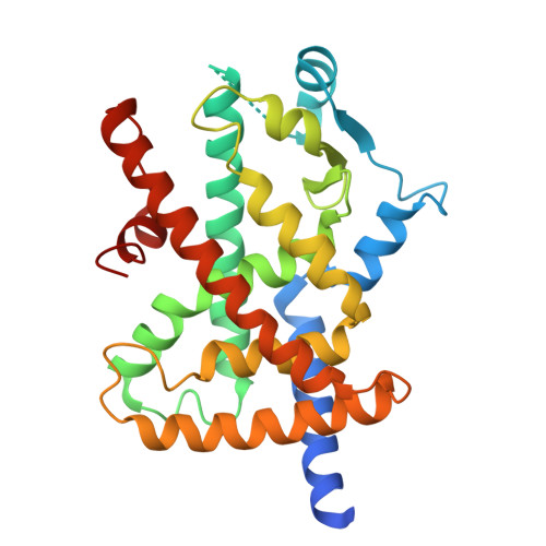
9HX2 - PubMed Abstract:
Insulin resistance (IR) is a pathological condition in which tissues exhibit a reduced response to normal or elevated levels of insulin. Type 2 diabetes mellitus (T2DM) and Metabolic Syndrome are the most prevalent disorders associated with IR. Most of the glitazones, traditional anti-diabetic drugs acting as Peroxisome Proliferator-Activated Receptor γ (PPARγ) agonists, have been withdrawn from the market. To mitigate the serious adverse effects associated with PPARγ agonism, a new opportunity is represented by the inhibitors of PPARγ phosphorylation by the Cyclin-Dependent Kinase 5 (CDK5). Their mechanism of action is mediated by the stabilization of the PPARγ β-sheet containing Ser245. Recently, we identified 4-(4-bromophenyl)-3-hydroxy-5-(3-hydroxyphenyl)furan-2(5H)-one (I) as a PPARγ non-agonist, capable of blocking the phosphorylation of the enzyme without direct effects on either CDK5 or PPARγ. Here, we isolated the two enantiomers of I, unambiguously defined their absolute configuration through single crystal X-ray diffraction and demonstrated by Grating-Coupled Interferometry binding assays that both (S)-I and (R)-I exhibited comparable affinity for PPARγ. Then, a library of 12 analogs was designed through structure-based modifications, optimizing the interactions within the ligand-binding domain. GCI analysis identified derivative 11, featuring an oxyacetic group in place of the initial hydroxyl function of the reference compound I, as the most promising candidate (K D = 186 nM). The crystal structure of the PPARγ-LBD/11 complex revealed a hydrogen bond interaction with Arg280, further stabilizing the binding conformation. These findings highlight the potential of γ-hydroxy lactone derivatives as PPARγ modulators and provide a foundation for future drug development targeting IR.
- Department of Pharmaceutical Sciences, University of Milan, via L. Mangiagalli 25, 20133, Milano, Italy; Department of Science and High Technology, University of Insubria, via Valleggio 9, 22100, Como, Italy.
Organizational Affiliation: