The bacterial ESCRT-III PspA rods thin lipid tubules and increase membrane curvature through helix alpha 0 interactions.
Hudina, E., Schott-Verdugo, S., Junglas, B., Kutzner, M., Ritter, I., Hellmann, N., Schneider, D., Gohlke, H., Sachse, C.(2025) Proc Natl Acad Sci U S A 122: e2506286122-e2506286122
- PubMed: 40758888
- DOI: https://doi.org/10.1073/pnas.2506286122
- Primary Citation of Related Structures:
9HZM, 9HZN, 9HZO, 9HZP, 9HZQ, 9HZR, 9HZS, 9HZT, 9HZU, 9HZV, 9HZW, 9HZX, 9HZY, 9HZZ, 9I00, 9I01 - PubMed Abstract:
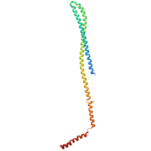
The phage shock protein A (PspA), a bacterial member of the endosomal sorting complexes required for transport (ESCRT)-III superfamily, forms rod-shaped helical assemblies that internalize membrane tubules. The N-terminal helix α0 of PspA (and other ESCRT-III members) has been suggested to act as a membrane anchor; the detailed mechanism, however, of how it binds to membranes and eventually triggers membrane fusion and/or fission events remains unclear. By solving a total of 15 cryoelectron microscopy (cryo-EM) structures of PspA and a truncation lacking the N-terminal helix α0 in the presence of Escherichia coli polar lipid membranes, we show in molecular detail how PspA interacts with and remodels membranes: Binding of the N-terminal helix α0 in the outer tubular membrane leaflet induces membrane curvature, supporting membrane tubulation by PspA. Detailed molecular dynamics simulations and free energy computations of interactions between the helix α0 and negatively charged membranes suggest a compensating mechanism between helix-membrane interactions and the energy contributions required for membrane bending. The energetic considerations are in line with the membrane structures observed in the cryo-EM images of tubulated membrane vesicles, fragmented vesicles inside tapered PspA rods, and shedded vesicles emerging at the thinner PspA rod ends. Our results provide insights into the molecular determinants and a potential mechanism of vesicular membrane remodeling mediated by a member of the ESCRT-III superfamily.
- Ernst-Ruska Centre for Microscopy and Spectroscopy with Electrons, ER-C-3: Structural Biology, Forschungszentrum Jülich, Jülich 52425, Germany.
Organizational Affiliation: