9ITB | pdb_00009itb

LPA-bound LPAR6 in complex with miniGq


Experimental Data Snapshot

  • Method: ELECTRON MICROSCOPY
  • Resolution: 2.89 Å
  • Aggregation State: PARTICLE 
  • Reconstruction Method: SINGLE PARTICLE 

wwPDB Validation   3D Report Full Report


This is version 1.2 of the entry. See complete history


Literature

Molecular mechanism of ligand recognition and activation of lysophosphatidic acid receptor LPAR6.

Duan, Y.Xu, Z.Hao, B.Zhang, A.Guo, C.He, Y.

(2025) Proc Natl Acad Sci U S A 122: e2415426122-e2415426122

  • DOI: https://doi.org/10.1073/pnas.2415426122
  • Primary Citation of Related Structures:  
    9ITB, 9ITE

  • PubMed Abstract: 

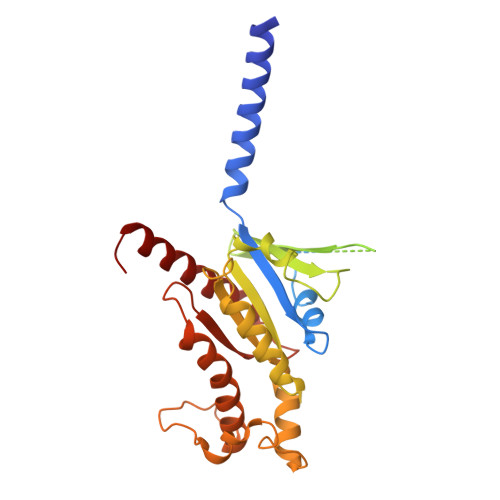
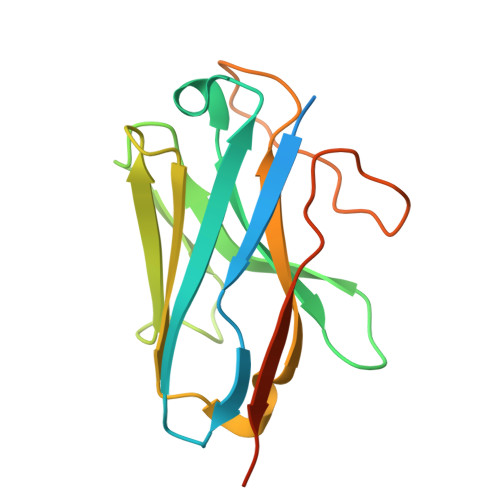
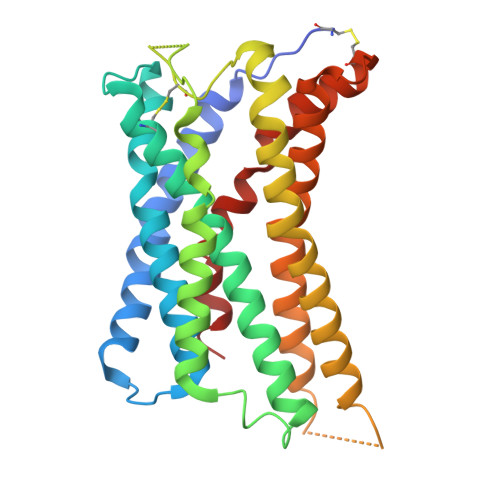
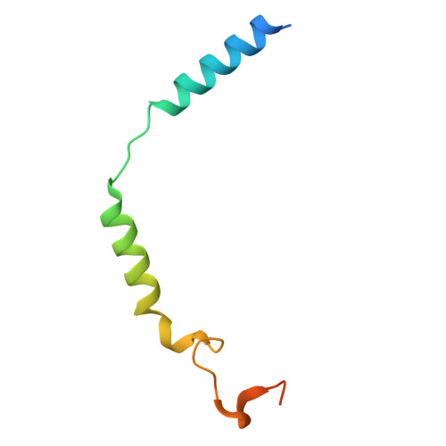
    Lysophosphatidic acid (LPA) exerts its physiological roles through the endothelialdifferentiation gene (EDG) family LPA receptors (LPAR1-3) or the non-EDG family LPA receptors (LPAR4-6). LPAR6 plays crucial roles in hair loss and cancer progression, yet its structural information is very limited. Here, we report the cryoelectron microscopy structure of LPA-bound human LPAR6 in complex with a mini G 13 or G q protein. These structures reveal a distinct ligand binding and recognition mode that differs significantly from that of LPAR1. Specifically, LPA uses its charged head to form an extensive polar interaction network with key polar residues on the extracellular side of transmembrane helix 5-6 and the extracellular loop 2. Structural comparisons and homology analysis suggest that the EDG and non-EDG families use two distinct modes for LPA binding. The structural observations are validated through functional mutagenesis studies. We further uncover the mechanisms of LPAR6 activation and principles of G-protein coupling. The structural information revealed by our study lays the groundwork for understanding LPAR6 signaling and provides a rational basis for designing compounds targeting LPAR6.


  • Organizational Affiliation
    • Faculty of Life Sciences and Medicine, Harbin Institute of Technology Center for Life Sciences, School of Life Science and Technology, Harbin Institute of Technology, Harbin 150001, China.

Macromolecules
Find similar proteins by:  (by identity cutoff)  |  3D Structure
Entity ID: 1
MoleculeChains Sequence LengthOrganismDetailsImage
engineered miniGaq362Homo sapiensMutation(s): 0 
Entity Groups  
Sequence Clusters30% Identity50% Identity70% Identity90% Identity95% Identity100% Identity
Sequence Annotations
Expand
  • Reference Sequence
Find similar proteins by:  (by identity cutoff)  |  3D Structure
Entity ID: 2
MoleculeChains Sequence LengthOrganismDetailsImage
Guanine nucleotide-binding protein G(I)/G(S)/G(T) subunit beta-1345Homo sapiensMutation(s): 0 
Gene Names: GNB1
UniProt & NIH Common Fund Data Resources
Find proteins for P62873 (Homo sapiens)
Explore P62873 
Go to UniProtKB:  P62873
PHAROS:  P62873
GTEx:  ENSG00000078369 
Entity Groups  
Sequence Clusters30% Identity50% Identity70% Identity90% Identity95% Identity100% Identity
UniProt GroupP62873
Sequence Annotations
Expand
  • Reference Sequence
Find similar proteins by:  (by identity cutoff)  |  3D Structure
Entity ID: 3
MoleculeChains Sequence LengthOrganismDetailsImage
Nb35C [auth N]161Lama glamaMutation(s): 0 
Entity Groups  
Sequence Clusters30% Identity50% Identity70% Identity90% Identity95% Identity100% Identity
Sequence Annotations
Expand
  • Reference Sequence
Find similar proteins by:  (by identity cutoff)  |  3D Structure
Entity ID: 4
MoleculeChains Sequence LengthOrganismDetailsImage
Lysophosphatidic acid receptor 6D [auth R]294Homo sapiensMutation(s): 0 
Gene Names: LPAR6P2RY5
Membrane Entity: Yes 
UniProt & NIH Common Fund Data Resources
Find proteins for P43657 (Homo sapiens)
Explore P43657 
Go to UniProtKB:  P43657
PHAROS:  P43657
GTEx:  ENSG00000139679 
Entity Groups  
Sequence Clusters30% Identity50% Identity70% Identity90% Identity95% Identity100% Identity
UniProt GroupP43657
Sequence Annotations
Expand
  • Reference Sequence
Find similar proteins by:  (by identity cutoff)  |  3D Structure
Entity ID: 5
MoleculeChains Sequence LengthOrganismDetailsImage
Guanine nucleotide-binding protein G(I)/G(S)/G(O) subunit gamma-2E [auth G]71Homo sapiensMutation(s): 0 
Gene Names: GNG2
Membrane Entity: Yes 
UniProt & NIH Common Fund Data Resources
Find proteins for P59768 (Homo sapiens)
Explore P59768 
Go to UniProtKB:  P59768
PHAROS:  P59768
GTEx:  ENSG00000186469 
Entity Groups  
Sequence Clusters30% Identity50% Identity70% Identity90% Identity95% Identity100% Identity
UniProt GroupP59768
Sequence Annotations
Expand
  • Reference Sequence
Experimental Data & Validation

Experimental Data

  • Method: ELECTRON MICROSCOPY
  • Resolution: 2.89 Å
  • Aggregation State: PARTICLE 
  • Reconstruction Method: SINGLE PARTICLE 
EM Software:
TaskSoftware PackageVersion
MODEL REFINEMENTPHENIX

Structure Validation

View Full Validation Report


Entry History & Funding Information

Deposition Data


Funding OrganizationLocationGrant Number
National Natural Science Foundation of China (NSFC)China32070048

Revision History  (Full details and data files)

  • Version 1.0: 2025-01-15
    Type: Initial release
  • Version 1.1: 2025-02-05
    Changes: Data collection, Database references
  • Version 1.2: 2025-07-23
    Changes: Data collection