9IVK | pdb_00009ivk

cryo-EM structure of a tmFAP


Experimental Data Snapshot

  • Method: ELECTRON MICROSCOPY
  • Resolution: 2.74 Å
  • Aggregation State: PARTICLE 
  • Reconstruction Method: SINGLE PARTICLE 

wwPDB Validation   3D Report Full Report


This is version 1.3 of the entry. See complete history


Literature

De novo design of transmembrane fluorescence-activating proteins.

Zhu, J.Liang, M.Sun, K.Wei, Y.Guo, R.Zhang, L.Shi, J.Ma, D.Hu, Q.Huang, G.Lu, P.

(2025) Nature 640: 249-257

  • DOI: https://doi.org/10.1038/s41586-025-08598-8
  • Primary Citation of Related Structures:  
    8W6E, 8W6F, 9IVK

  • PubMed Abstract: 

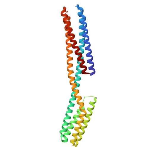
    The recognition of ligands by transmembrane proteins is essential for the exchange of materials, energy and information across biological membranes. Progress has been made in the de novo design of transmembrane proteins 1-6 , as well as in designing water-soluble proteins to bind small molecules 7-12 , but de novo design of transmembrane proteins that tightly and specifically bind to small molecules remains an outstanding challenge 13 . Here we present the accurate design of ligand-binding transmembrane proteins by integrating deep learning and energy-based methods. We designed pre-organized ligand-binding pockets in high-quality four-helix backbones for a fluorogenic ligand, and generated a transmembrane span using gradient-guided hallucination. The designer transmembrane proteins specifically activated fluorescence of the target fluorophore with mid-nanomolar affinity, exhibiting higher brightness and quantum yield compared to those of enhanced green fluorescent protein. These proteins were highly active in the membrane fraction of live bacterial and eukaryotic cells following expression. The crystal and cryogenic electron microscopy structures of the designer protein-ligand complexes were very close to the structures of the design models. We showed that the interactions between ligands and transmembrane proteins within the membrane can be accurately designed. Our work paves the way for the creation of new functional transmembrane proteins, with a wide range of applications including imaging, ligand sensing and membrane transport.


  • Organizational Affiliation

    College of Life Sciences, Zhejiang University, Hangzhou, China.


Macromolecules
Find similar proteins by:  (by identity cutoff)  |  3D Structure
Entity ID: 1
MoleculeChains Sequence LengthOrganismDetailsImage
HBC599 membrane protein binder285artificial sequencesMutation(s): 0 
Membrane Entity: Yes 
UniProt
Find proteins for P0ABE7 (Escherichia coli)
Explore P0ABE7 
Go to UniProtKB:  P0ABE7
Entity Groups  
Sequence Clusters30% Identity50% Identity70% Identity90% Identity95% Identity100% Identity
UniProt GroupP0ABE7
Sequence Annotations
Expand
  • Reference Sequence
Find similar proteins by:  (by identity cutoff)  |  3D Structure
Entity ID: 2
MoleculeChains Sequence LengthOrganismDetailsImage
Heavy chain, Fab fragmentB [auth H]229Homo sapiensMutation(s): 0 
Entity Groups  
Sequence Clusters30% Identity50% Identity70% Identity90% Identity95% Identity100% Identity
Sequence Annotations
Expand
  • Reference Sequence
Find similar proteins by:  (by identity cutoff)  |  3D Structure
Entity ID: 3
MoleculeChains Sequence LengthOrganismDetailsImage
Light Chain, Fab fragmentC [auth L]214Homo sapiensMutation(s): 0 
Entity Groups  
Sequence Clusters30% Identity50% Identity70% Identity90% Identity95% Identity100% Identity
Sequence Annotations
Expand
  • Reference Sequence
Small Molecules
Ligands 1 Unique
IDChains Name / Formula / InChI Key2D Diagram3D Interactions
KY6
Query on KY6

Download Ideal Coordinates CCD File 
D [auth A]4-[(Z)-1-cyano-2-{6-[(2-hydroxyethyl)(methyl)amino]-1-benzothiophen-2-yl}ethenyl]benzonitrile
C21 H17 N3 O S
WXTHEUULEXARCU-WOJGMQOQSA-N
Experimental Data & Validation

Experimental Data

  • Method: ELECTRON MICROSCOPY
  • Resolution: 2.74 Å
  • Aggregation State: PARTICLE 
  • Reconstruction Method: SINGLE PARTICLE 

Structure Validation

View Full Validation Report


Entry History & Funding Information

Deposition Data


Funding OrganizationLocationGrant Number
Ministry of Science and Technology (MoST, China)China2020YFA0909200

Revision History  (Full details and data files)

  • Version 1.0: 2024-11-20
    Type: Initial release
  • Version 1.1: 2025-02-26
    Changes: Data collection, Database references
  • Version 1.2: 2025-03-05
    Changes: Data collection, Database references
  • Version 1.3: 2025-04-16
    Changes: Data collection, Database references