


Structure of glutamine synthetase from ESKAPE pathogen Acinetobacter baumannii
Chandrasekaran, P., Killivalavan, A., Vaigundan, D., Yuvaraj, I., Manju, B., Sekar, K.To be published.
Experimental Data Snapshot
Starting Model: experimental
View more details
Entity ID: 1 | |||||
|---|---|---|---|---|---|
| Molecule | Chains | Sequence Length | Organism | Details | Image |
| Glutamine synthetase | 468 | Acinetobacter baumannii AB0057 | Mutation(s): 0 Gene Names: AB57_2783 EC: 6.3.1.2 | 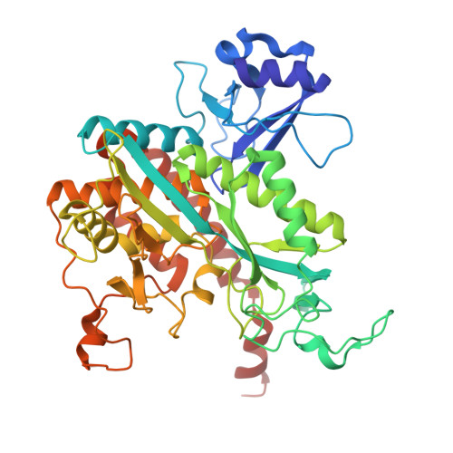 | |
UniProt | |||||
Find proteins for A0A7U3Y893 (Acinetobacter baumannii (strain AB0057)) Explore A0A7U3Y893 Go to UniProtKB: A0A7U3Y893 | |||||
Entity Groups | |||||
| Sequence Clusters | 30% Identity50% Identity70% Identity90% Identity95% Identity100% Identity | ||||
| UniProt Group | A0A7U3Y893 | ||||
Sequence AnnotationsExpand | |||||
| |||||
| Ligands 4 Unique | |||||
|---|---|---|---|---|---|
| ID | Chains | Name / Formula / InChI Key | 2D Diagram | 3D Interactions | |
| PGE (Subject of Investigation/LOI) Query on PGE | G [auth A] | TRIETHYLENE GLYCOL C6 H14 O4 ZIBGPFATKBEMQZ-UHFFFAOYSA-N |  | ||
| GOL (Subject of Investigation/LOI) Query on GOL | H [auth A] I [auth A] L [auth B] M [auth B] P [auth C] | GLYCEROL C3 H8 O3 PEDCQBHIVMGVHV-UHFFFAOYSA-N |  | ||
| EDO (Subject of Investigation/LOI) Query on EDO | Y [auth F] | 1,2-ETHANEDIOL C2 H6 O2 LYCAIKOWRPUZTN-UHFFFAOYSA-N |  | ||
| MG (Subject of Investigation/LOI) Query on MG | AA [auth F] J [auth A] K [auth A] N [auth B] O [auth B] | MAGNESIUM ION Mg JLVVSXFLKOJNIY-UHFFFAOYSA-N |  | ||
| Length ( Å ) | Angle ( ˚ ) |
|---|---|
| a = 260.909 | α = 90 |
| b = 260.909 | β = 90 |
| c = 154.18 | γ = 90 |
| Software Name | Purpose |
|---|---|
| REFMAC | refinement |
| autoPX | data processing |
| iMOSFLM | data reduction |
| Aimless | data scaling |
| MOLREP | phasing |
| Funding Organization | Location | Grant Number |
|---|---|---|
| Department of Science & Technology (DST, India) | India | Meit/R&D/HPC/2(1)/2014 |


