9KTI | pdb_00009kti

CryoEM structure of a 2,3-hydroxycinnamic acid 1,2-dioxygenase MhpB in substrate bound form


Experimental Data Snapshot

  • Method: ELECTRON MICROSCOPY
  • Resolution: 2.59 Å
  • Aggregation State: PARTICLE 
  • Reconstruction Method: SINGLE PARTICLE 

wwPDB Validation   3D Report Full Report


This is version 1.0 of the entry. See complete history


Literature

Structural and catalytic insights into MhpB: A dioxygenase enzyme for degrading catecholic pollutants.

Dong, X.Xu, M.Wu, M.Wang, Y.Cheng, X.Jiang, W.Zheng, D.Omar, A.H.Cheng, Y.Li, A.Ma, L.Xing, Q.

(2025) J Hazard Mater 488: 137431-137431

  • DOI: https://doi.org/10.1016/j.jhazmat.2025.137431
  • Primary Citation of Related Structures:  
    8K04, 9KTI

  • PubMed Abstract: 

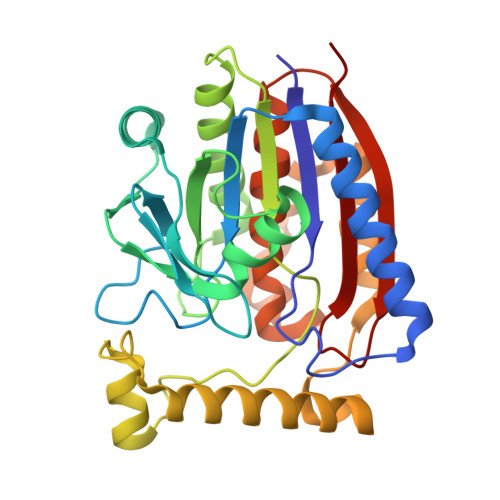
    The increasing environmental pollution from persistent aromatic compounds requires effective biodegradation strategies. In this study, we focused on MhpB, an extradiol dioxygenase (EDO) from Escherichia coli. It is known for its role in the degradation of catechols, key intermediates in the degradation of aromatic compounds. We report the high-resolution structure of MhpB determined by cryo-electron microscopy, revealing a decameric conformation with the catalytic chamber at the side. The structure-based analysis allowed us to investigate the substrate-enzyme interaction and the substrate selectivity, which are crucial for its catalytic function. Site-directed mutagenesis was used to modulate the in vitro and in vivo substrate preference of MhpB, enhancing its potential for industrial applications in pollutant degradation. The study provides insight into the mechanism of the enzyme and paves the way for the development of engineered EDOs for environmental remediation of aromatic pollutants.


  • Organizational Affiliation

    State Key Laboratory of Biocatalysis and Enzyme Engineering, College of Life Sciences, Hubei University, Wuhan 430074, China. Electronic address: dongxu@hubu.edu.cn.


Macromolecules
Find similar proteins by:  (by identity cutoff)  |  3D Structure
Entity ID: 1
MoleculeChains Sequence LengthOrganismDetailsImage
2,3-dihydroxyphenylpropionate/2,3-dihydroxicinnamic acid 1,2-dioxygenase
A, B, C, D, E
A, B, C, D, E, F, G, H, I, J
314Escherichia coli K-12Mutation(s): 0 
Gene Names: mhpBb0348JW0339
EC: 1.13.11.16
UniProt
Find proteins for P0ABR9 (Escherichia coli (strain K12))
Explore P0ABR9 
Go to UniProtKB:  P0ABR9
Entity Groups  
Sequence Clusters30% Identity50% Identity70% Identity90% Identity95% Identity100% Identity
UniProt GroupP0ABR9
Sequence Annotations
Expand
  • Reference Sequence
Small Molecules
Ligands 2 Unique
IDChains Name / Formula / InChI Key2D Diagram3D Interactions
A1EG2 (Subject of Investigation/LOI)
Query on A1EG2

Download Ideal Coordinates CCD File 
BA [auth I]
DA [auth J]
L [auth A]
N [auth B]
O [auth C]
BA [auth I],
DA [auth J],
L [auth A],
N [auth B],
O [auth C],
R [auth D],
T [auth E],
V [auth F],
X [auth G],
Z [auth H]
3-[2,3-bis(oxidanyl)phenyl]propanoic acid
C9 H10 O4
QZDSXQJWBGMRLU-UHFFFAOYSA-N
FE2
Query on FE2

Download Ideal Coordinates CCD File 
AA [auth I]
CA [auth J]
K [auth A]
M [auth B]
P [auth C]
AA [auth I],
CA [auth J],
K [auth A],
M [auth B],
P [auth C],
Q [auth D],
S [auth E],
U [auth F],
W [auth G],
Y [auth H]
FE (II) ION
Fe
CWYNVVGOOAEACU-UHFFFAOYSA-N
Experimental Data & Validation

Experimental Data

  • Method: ELECTRON MICROSCOPY
  • Resolution: 2.59 Å
  • Aggregation State: PARTICLE 
  • Reconstruction Method: SINGLE PARTICLE 

Structure Validation

View Full Validation Report


Entry History & Funding Information

Deposition Data


Funding OrganizationLocationGrant Number
National Science Foundation (NSF, China)China32371277
National Science Foundation (NSF, China)China32301028

Revision History  (Full details and data files)

  • Version 1.0: 2025-02-12
    Type: Initial release