9LO2 | pdb_00009lo2

Crystal Structure of Anti-CRISPR protein


Experimental Data Snapshot

  • Method: X-RAY DIFFRACTION
  • Resolution: 2.29 Å
  • R-Value Free: 
    0.241 (Depositor), 0.239 (DCC) 
  • R-Value Work: 
    0.208 (Depositor), 0.215 (DCC) 
  • R-Value Observed: 
    0.211 (Depositor) 

wwPDB Validation   3D Report Full Report


This is version 1.1 of the entry. See complete history


Literature

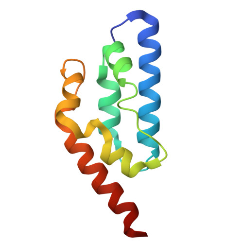
Structural Investigation of the Anti-CRISPR Protein AcrIE7.

Kang, J.Park, C.Lee, G.Koo, J.Oh, H.Kim, E.H.Bae, E.Suh, J.Y.

(2025) Proteins 93: 1645-1656

  • DOI: https://doi.org/10.1002/prot.26832
  • Primary Citation of Related Structures:  
    9LO1, 9LO2

  • PubMed Abstract: 

    The CRISPR-Cas system is an adaptive immune system in prokaryotes that provides protection against bacteriophages. As a countermeasure, bacteriophages have evolved various anti-CRISPR proteins that neutralize CRISPR-Cas immunity. Here, we report the structural and functional investigation of AcrIE7, which inhibits the type I-E CRISPR-Cas system in Pseudomonas aeruginosa. We determined both crystal and solution structures of AcrIE7, which revealed a novel helical fold. In binding assays using various biochemical methods, AcrIE7 did not tightly interact with a single Cas component in the type I-E Cascade complex or the CRISPR adaptation machinery. In contrast, AlphaFold modeling with our experimentally determined AcrIE7 structure predicted that AcrIE7 interacts with Cas3 in the type I-E CRISPR-Cas system in P. aeruginosa. Our findings are consistent with a model where AcrIE7 inhibits Cas3 and also highlight the effectiveness and limitations of AlphaFold modeling.


  • Organizational Affiliation
    • Department of Agricultural Biotechnology, Seoul National University, Seoul, Korea.

Macromolecules
Find similar proteins by:  (by identity cutoff)  |  3D Structure
Entity ID: 1
MoleculeChains Sequence LengthOrganismDetailsImage
Anti-CRISPR protein, AcrIE7
A, B, C
107Pseudomonas aeruginosaMutation(s): 0 
Entity Groups  
Sequence Clusters30% Identity50% Identity70% Identity90% Identity95% Identity100% Identity
Sequence Annotations
Expand
  • Reference Sequence
Experimental Data & Validation

Experimental Data

  • Method: X-RAY DIFFRACTION
  • Resolution: 2.29 Å
  • R-Value Free:  0.241 (Depositor), 0.239 (DCC) 
  • R-Value Work:  0.208 (Depositor), 0.215 (DCC) 
  • R-Value Observed: 0.211 (Depositor) 
Space Group: P 63 2 2
Unit Cell:
Length ( Å )Angle ( ˚ )
a = 109.203α = 90
b = 109.203β = 90
c = 187.956γ = 120
Software Package:
Software NamePurpose
HKL-2000data collection
HKL-2000data scaling
PHENIXphasing
PHENIXmodel building
PHENIXrefinement

Structure Validation

View Full Validation Report


Entry History & Funding Information

Deposition Data


Funding OrganizationLocationGrant Number
National Research Foundation (NRF, Korea)Korea, Republic OfNRF-2022R1A2C1009804
National Research Foundation (NRF, Korea)Korea, Republic OfRS-2023-00207820

Revision History  (Full details and data files)

  • Version 1.0: 2025-06-18
    Type: Initial release
  • Version 1.1: 2025-08-13
    Changes: Database references