9M7M | pdb_00009m7m

Crystal structure of human pseudouridine 5'-monophosphate phosphatase (hHDHD1A) complexed with pseudouridine


Experimental Data Snapshot

  • Method: X-RAY DIFFRACTION
  • Resolution: 1.26 Å
  • R-Value Free: 
    0.209 (Depositor), 0.212 (DCC) 
  • R-Value Work: 
    0.184 (Depositor), 0.186 (DCC) 
  • R-Value Observed: 
    0.184 (Depositor) 

wwPDB Validation   3D Report Full Report


Ligand Structure Quality Assessment 


This is version 1.1 of the entry. See complete history


Literature

Structural and functional insights into the substrate specificity of the pseudouridine monophosphate phosphatase HDHD1A.

Seo, S.Kim, M.Rhee, S.

(2025) J Biological Chem 301: 110257-110257

  • DOI: https://doi.org/10.1016/j.jbc.2025.110257
  • Primary Citation of Related Structures:  
    9M7L, 9M7M

  • PubMed Abstract: 

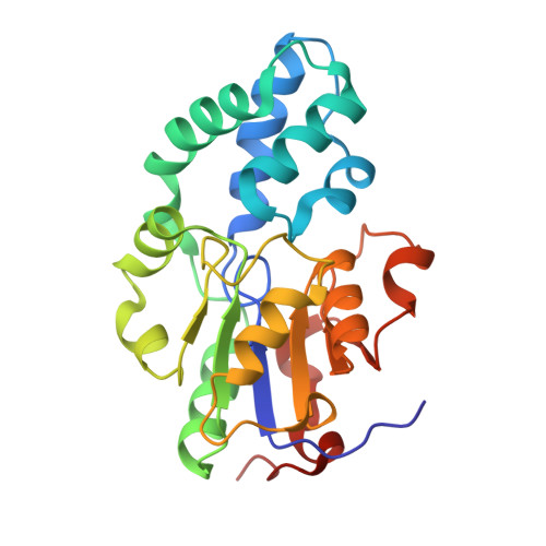
    Pseudouridine (Ψ) is one of the most abundant RNA modifications. Following RNA degradation, Ψ nucleotides are dephosphorylated and catabolized into uracil and ribose 5'-phosphate via a two-step enzymatic reaction catalyzed by enzymes present in many bacteria and eukaryotes, but not in mammals. Malfunction of Ψ catabolism has adverse physiological effects in plants. In humans, the enzyme HDHD1A dephosphorylates pseudouridine 5'-monophosphate (ΨMP), and the resulting Ψ is excreted in the urine. In this study, we determined the crystal structures of human HDHD1A (hHDHD1A) complexed with Ψ. The structure of hHDHD1A consists of a catalytic domain with a Rossmann α/β-fold and a cap domain, with a magnesium ion at the junction of the two domains. Ψ is bound to the active site in an orientation where its nucleobase, uracil-Ψ, is surrounded by the cap domain residues, and the ribose moiety is located next to the Mg 2+ -binding site. The active site is composed mainly of hydrophobic residues, but two essential charged residues, Glu23 and Lys46, are present in the vicinity of uracil-Ψ. Glu23 interacts with the Ψ-specific N1 atom, while Lys46 interacts with the O2 atom of uracil-Ψ. Mutagenesis and kinetic analysis indicated that active site residues are involved in substrate binding and/or catalysis. In addition to Ψ-specific hydrophilic interactions, shape complementarity between ΨMP and the active site pocket is a key element underlying substrate specificity in hHDHD1A. This study provided structural and functional insights into the substrate specificity of hHDHD1A for ΨMP, highlighting both similarities and differences compared to other Ψ-recognizing enzymes.


  • Organizational Affiliation
    • Department of Agricultural Biotechnology, Seoul National University, Seoul, Korea.

Macromolecules
Find similar proteins by:  (by identity cutoff)  |  3D Structure
Entity ID: 1
MoleculeChains Sequence LengthOrganismDetailsImage
Pseudouridine-5'-phosphatase226Homo sapiensMutation(s): 0 
Gene Names: PUDPDXF68S1EFAM16AXGS1HDHD1HDHD1A
EC: 3.1.3.96
UniProt & NIH Common Fund Data Resources
Find proteins for Q08623 (Homo sapiens)
Explore Q08623 
Go to UniProtKB:  Q08623
PHAROS:  Q08623
GTEx:  ENSG00000130021 
Entity Groups  
Sequence Clusters30% Identity50% Identity70% Identity90% Identity95% Identity100% Identity
UniProt GroupQ08623
Sequence Annotations
Expand
  • Reference Sequence
Small Molecules
Ligands 2 Unique
IDChains Name / Formula / InChI Key2D Diagram3D Interactions
FJF (Subject of Investigation/LOI)
Query on FJF

Download Ideal Coordinates CCD File 
C [auth A]5-[(2~{S},3~{R},4~{S},5~{R})-5-(hydroxymethyl)-3,4-bis(oxidanyl)oxolan-2-yl]-1~{H}-pyrimidine-2,4-dione
C9 H12 N2 O6
PTJWIQPHWPFNBW-GBNDHIKLSA-N
MG (Subject of Investigation/LOI)
Query on MG

Download Ideal Coordinates CCD File 
B [auth A]MAGNESIUM ION
Mg
JLVVSXFLKOJNIY-UHFFFAOYSA-N
Experimental Data & Validation

Experimental Data

  • Method: X-RAY DIFFRACTION
  • Resolution: 1.26 Å
  • R-Value Free:  0.209 (Depositor), 0.212 (DCC) 
  • R-Value Work:  0.184 (Depositor), 0.186 (DCC) 
  • R-Value Observed: 0.184 (Depositor) 
Space Group: P 2 21 21
Unit Cell:
Length ( Å )Angle ( ˚ )
a = 51.62α = 90
b = 61.09β = 90
c = 68.25γ = 90
Software Package:
Software NamePurpose
PHENIXrefinement
XDSdata reduction
XDSdata scaling
PHENIXphasing

Structure Validation

View Full Validation Report


Ligand Structure Quality Assessment 


Entry History & Funding Information

Deposition Data


Funding OrganizationLocationGrant Number
National Research Foundation (NRF, Korea)Korea, Republic Of--

Revision History  (Full details and data files)

  • Version 1.0: 2025-06-18
    Type: Initial release
  • Version 1.1: 2025-07-02
    Changes: Database references