9NE6 | pdb_00009ne6

Human polymerase epsilon bound to PCNA and DNA with an in-situ-generated mismatch in the mismatch-editing state


Experimental Data Snapshot

  • Method: ELECTRON MICROSCOPY
  • Resolution: 3.11 Å
  • Aggregation State: PARTICLE 
  • Reconstruction Method: SINGLE PARTICLE 

wwPDB Validation   3D Report Full Report


This is version 1.0 of the entry. See complete history


Literature

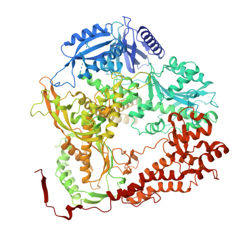
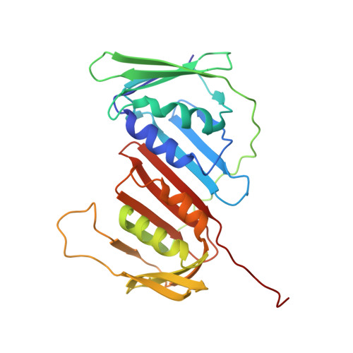
The proofreading mechanism of the human leading-strand DNA polymerase epsilon holoenzyme.

Wang, F.He, Q.O'Donnell, M.E.Li, H.

(2025) Proc Natl Acad Sci U S A 122: e2507232122-e2507232122

  • DOI: https://doi.org/10.1073/pnas.2507232122
  • Primary Citation of Related Structures:  
    9NE6, 9NE7, 9NE8, 9NE9, 9NEA

  • PubMed Abstract: 

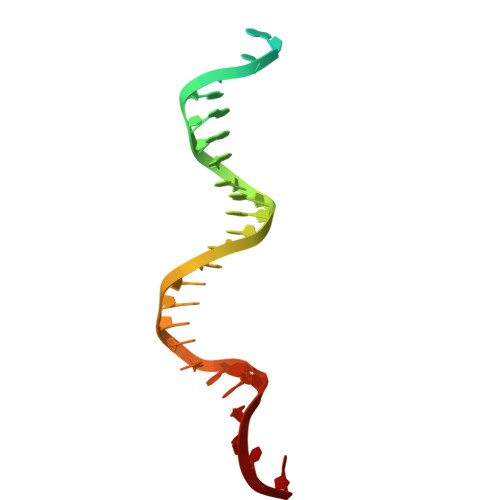
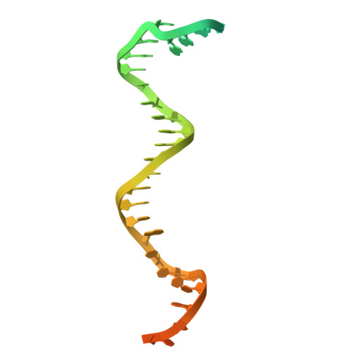
    The eukaryotic leading-strand DNA polymerase ε (Polε) is a dual-function enzyme with a proofreading 3'-5' exonuclease ( exo ) site located 40 Å from the DNA synthesizing pol site. Errors in Polε proofreading can cause various mutations, including C-to-G transversions, the most prevalent mutation in cancers and genetic diseases. Polε interacts with all three subunits of the PCNA ring to assemble a functional holoenzyme. Despite previous studies on proofreading of several Pol's, how Polε-or any Pol complexed with its sliding clamp-proofreads a mismatch generated in situ has been unknown. We show here by cryo-EM that a template/primer DNA substrate with a preexisting mismatch cannot enter the exo site of Polε-PCNA holoenzyme, but a mismatch generated in situ in the pol site yields three bona fide proofreading intermediates of Polε-PCNA holoenzyme. These intermediates reveal how the mismatch is dislodged from the pol site, how the DNA unwinds six base pairs, and how the unpaired primer 3'-end is inserted into the exo site for cleavage. These results unexpectedly demonstrate that PCNA imposes strong steric constraints that extend unwinding and direct the trajectory of mismatched DNA and that this trajectory is dramatically different than for Polε in the absence of PCNA. These findings suggest a physiologically relevant proofreading mechanism for the human Polε holoenzyme.


  • Organizational Affiliation

    Department of Structural Biology, Van Andel Institute, Grand Rapids, MI 49503.


Macromolecules

Find similar proteins by:  (by identity cutoff)  |  3D Structure
Entity ID: 1
MoleculeChains Sequence LengthOrganismDetailsImage
DNA polymerase epsilon catalytic subunit A1,200Homo sapiensMutation(s): 0 
Gene Names: POLEPOLE1
EC: 2.7.7.7 (PDB Primary Data), 3.1.11 (PDB Primary Data)
UniProt & NIH Common Fund Data Resources
Find proteins for Q07864 (Homo sapiens)
Explore Q07864 
Go to UniProtKB:  Q07864
PHAROS:  Q07864
GTEx:  ENSG00000177084 
Entity Groups  
Sequence Clusters30% Identity50% Identity70% Identity90% Identity95% Identity100% Identity
UniProt GroupQ07864
Sequence Annotations
Expand
  • Reference Sequence
Find similar proteins by:  (by identity cutoff)  |  3D Structure
Entity ID: 2
MoleculeChains Sequence LengthOrganismDetailsImage
Proliferating cell nuclear antigen
B, C, D
261Homo sapiensMutation(s): 0 
Gene Names: PCNA
UniProt & NIH Common Fund Data Resources
Find proteins for P12004 (Homo sapiens)
Explore P12004 
Go to UniProtKB:  P12004
PHAROS:  P12004
GTEx:  ENSG00000132646 
Entity Groups  
Sequence Clusters30% Identity50% Identity70% Identity90% Identity95% Identity100% Identity
UniProt GroupP12004
Sequence Annotations
Expand
  • Reference Sequence
Find similar nucleic acids by:  (by identity cutoff)  |  3D Structure
Entity ID: 3
MoleculeChains LengthOrganismImage
DNA (33-MER)E [auth P]33synthetic construct
Sequence Annotations
Expand
  • Reference Sequence
Find similar nucleic acids by:  (by identity cutoff)  |  3D Structure
Entity ID: 4
MoleculeChains LengthOrganismImage
DNA (47-MER)F [auth T]47synthetic construct
Sequence Annotations
Expand
  • Reference Sequence
Small Molecules
Ligands 1 Unique
IDChains Name / Formula / InChI Key2D Diagram3D Interactions
SF4 (Subject of Investigation/LOI)
Query on SF4

Download Ideal Coordinates CCD File 
G [auth A]IRON/SULFUR CLUSTER
Fe4 S4
LJBDFODJNLIPKO-UHFFFAOYSA-N
Experimental Data & Validation

Experimental Data

  • Method: ELECTRON MICROSCOPY
  • Resolution: 3.11 Å
  • Aggregation State: PARTICLE 
  • Reconstruction Method: SINGLE PARTICLE 
EM Software:
TaskSoftware PackageVersion
MODEL REFINEMENTPHENIX

Structure Validation

View Full Validation Report


Entry History & Funding Information

Deposition Data


Funding OrganizationLocationGrant Number
National Institutes of Health/National Institute of General Medical Sciences (NIH/NIGMS)United StatesGM131754
National Institutes of Health/National Institute of General Medical Sciences (NIH/NIGMS)United StatesGM148159

Revision History  (Full details and data files)

  • Version 1.0: 2025-06-04
    Type: Initial release