9NH3 | pdb_00009nh3

Helicobacter pylori strain SS1 KatA


Experimental Data Snapshot

  • Method: X-RAY DIFFRACTION
  • Resolution: 1.87 Å
  • R-Value Free: 
    0.191 (Depositor), 0.191 (DCC) 
  • R-Value Work: 
    0.142 (Depositor), 0.141 (DCC) 
  • R-Value Observed: 
    0.143 (Depositor) 

Starting Model: experimental
View more details

wwPDB Validation   3D Report Full Report


Ligand Structure Quality Assessment 


This is version 1.0 of the entry. See complete history


Literature

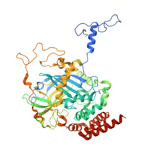
AlphaFold 3 accurately models natural variants of Helicobacter pylori catalase KatA.

Baylink, A.

(2025) bioRxiv 

  • DOI: https://doi.org/10.1101/2025.06.02.657526
  • Primary Citation of Related Structures:  
    9NH3

  • PubMed Abstract: 

    Subtle changes in protein sequence can equate to large changes in function, such as enabling pathogens to evade the immune system, hindering antibody recognition of antigens, or conferring antibiotic resistance. Even single amino acid substitutions may alter ligand binding affinity, enzymatic activity, and protein stability. Yet, due to limitations in time and resources, proteins closely related in sequence to those already characterized often remain unexamined. AlphaFold has emerged as a promising tool for protein structure prediction, though its utility in modeling single amino acid substitutions remains uncertain. In this study, we assessed AlphaFold 3's accuracy in modeling natural variants of the Helicobacter pylori catalase KatA by comparing its predictions to a novel high-resolution crystal structure of KatA from strain SS1. This variant contains key substitutions at residues 234, 237, 255, and 421 relative to the well-characterized strain 26695. AlphaFold 3 models accurately reproduced the global structure and local conformations of most variant residues, with high fidelity in conservative substitutions but variable accuracy in more flexible or interface-exposed sites. We further explored how user inputs, such as incorrect oligomeric states or sequence modifications, influence prediction quality. While AlphaFold 3 consistently produced high-quality models, deviations at variant sites occurred when incorrect oligomeric states were specified. Our findings highlight both the strengths and limitations of AlphaFold 3 in modeling natural protein variants and underscore the importance of accurate user input for reliable structural predictions.


  • Organizational Affiliation

    Department of Veterinary Microbiology and Pathology, Washington State University, Pullman, Washington, USA.


Macromolecules
Find similar proteins by:  (by identity cutoff)  |  3D Structure
Entity ID: 1
MoleculeChains Sequence LengthOrganismDetailsImage
Catalase
A, B
505Helicobacter pylori SS1Mutation(s): 0 
Gene Names: katA
EC: 1.11.1.6
UniProt
Find proteins for F4ZZ52 (Helicobacter pylori SS1)
Explore F4ZZ52 
Go to UniProtKB:  F4ZZ52
Entity Groups  
Sequence Clusters30% Identity50% Identity70% Identity90% Identity95% Identity100% Identity
UniProt GroupF4ZZ52
Sequence Annotations
Expand
  • Reference Sequence
Small Molecules
Ligands 4 Unique
IDChains Name / Formula / InChI Key2D Diagram3D Interactions
HEM (Subject of Investigation/LOI)
Query on HEM

Download Ideal Coordinates CCD File 
C [auth A],
H [auth B]
PROTOPORPHYRIN IX CONTAINING FE
C34 H32 Fe N4 O4
KABFMIBPWCXCRK-RGGAHWMASA-L
GOL
Query on GOL

Download Ideal Coordinates CCD File 
D [auth A],
J [auth B]
GLYCEROL
C3 H8 O3
PEDCQBHIVMGVHV-UHFFFAOYSA-N
CL
Query on CL

Download Ideal Coordinates CCD File 
I [auth B]CHLORIDE ION
Cl
VEXZGXHMUGYJMC-UHFFFAOYSA-M
NA
Query on NA

Download Ideal Coordinates CCD File 
E [auth A]
F [auth A]
G [auth A]
K [auth B]
L [auth B]
E [auth A],
F [auth A],
G [auth A],
K [auth B],
L [auth B],
M [auth B]
SODIUM ION
Na
FKNQFGJONOIPTF-UHFFFAOYSA-N
Experimental Data & Validation

Experimental Data

  • Method: X-RAY DIFFRACTION
  • Resolution: 1.87 Å
  • R-Value Free:  0.191 (Depositor), 0.191 (DCC) 
  • R-Value Work:  0.142 (Depositor), 0.141 (DCC) 
  • R-Value Observed: 0.143 (Depositor) 
Space Group: P 21 2 21
Unit Cell:
Length ( Å )Angle ( ˚ )
a = 67.512α = 90
b = 96.198β = 90
c = 154.754γ = 90
Software Package:
Software NamePurpose
PHENIXrefinement
Cootmodel building
Aimlessdata scaling
DIALSdata reduction
PHASERphasing

Structure Validation

View Full Validation Report


Ligand Structure Quality Assessment 


Entry History & Funding Information

Deposition Data

  • Released Date: 2025-07-23 
  • Deposition Author(s): Baylink, A.

Funding OrganizationLocationGrant Number
National Institutes of Health/National Institute Of Allergy and Infectious Diseases (NIH/NIAID)United States4R00AI148587-03

Revision History  (Full details and data files)

  • Version 1.0: 2025-07-23
    Type: Initial release