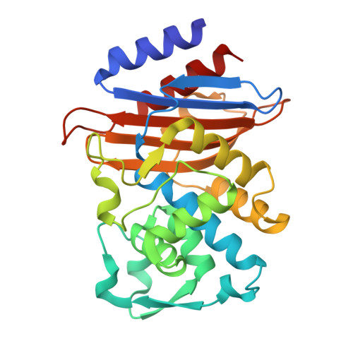
Combining MicroED and native mass spectrometry for structural discovery of enzyme-small molecule complexes.
Vlahakis, N.W., Flowers, C.W., Liu, M., Agdanowski, M.P., Johnson, S., Summers, J.A., Jacobs, L.M.C., Keyser, C., Russell, P., Rose, S.L., Orlans, J., Adhami, N., Chen, Y., Sawaya, M.R., Basu, S., de Sanctis, D., Chen, Y., Wakatsuki, S., Nelson, H.M., Loo, J.A., Tang, Y., Rodriguez, J.A.(2025) Proc Natl Acad Sci U S A 122: e2503780122-e2503780122
- PubMed: 40720654
- DOI: https://doi.org/10.1073/pnas.2503780122
- Primary Citation of Related Structures:
9NBP, 9NBQ, 9NC1, 9NCA, 9NCC, 9OQE, 9OR3, 9OR7, 9ORB, 9ORG, 9ORH, 9ORL, 9ORS, 9ORV, 9ORW, 9ORX, 9ORY, 9ORZ, 9OS0, 9OS1, 9OS8 - PubMed Abstract:
With the goal of accelerating the discovery of small molecule-protein complexes, we leverage fast, low-dose, event-based electron counting microcrystal electron diffraction (MicroED) data collection and native mass spectrometry. This approach, which we term electron diffraction with native mass spectrometry (ED-MS), allows assignment of protein target structures bound to ligands with data obtained from crystal slurries soaked with mixtures of known inhibitors and crude biosynthetic reactions. This extends to libraries of printed ligands dispensed directly onto TEM grids for later soaking with microcrystal slurries, and complexes with noncovalent ligands. ED-MS resolves structures of the natural product, epoxide-based cysteine protease inhibitor E-64, and its biosynthetic analogs bound to the model cysteine protease, papain. It further identifies papain binding to its preferred natural products, by showing that two analogs of E-64 outcompete others in binding to papain crystals, and by detecting papain bound to E-64 and an analog from crude biosynthetic reactions, without purification. ED-MS also resolves binding of the CTX-M-14 β-lactamase, a target of active drug development, to the non-β-lactam inhibitor, avibactam, alone or in a cocktail of unrelated compounds. These results illustrate the utility of ED-MS for natural product ligand discovery and for structure-based screening of small molecule binders to macromolecular targets, promising utility for drug discovery.
- Department of Chemistry and Biochemistry, University of California, Los Angeles, CA 90095.
Organizational Affiliation:


