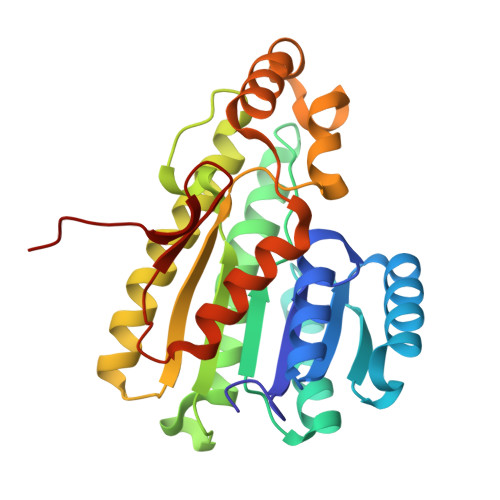
9PFM | pdb_00009pfm

Crystal structure of human 15-PGDH in complex with small molecule compound 8b


Experimental Data Snapshot

  • Method: X-RAY DIFFRACTION
  • Resolution: 1.88 Å
  • R-Value Free: 
    0.230 (Depositor), 0.232 (DCC) 
  • R-Value Work: 
    0.179 (Depositor), 0.180 (DCC) 
  • R-Value Observed: 
    0.182 (Depositor) 

Starting Model: experimental
View more details

wwPDB Validation   3D Report Full Report


Ligand Structure Quality Assessment 


This is version 1.1 of the entry. See complete history


Literature

Knowledge and Structure-Based Drug Design of 15-PGDH Inhibitors.

Dodda, L.S.Campos, S.Ciccone, D.Carreiro, S.Leit, S.Brennan, D.Zephyr, J.Jacques-O'Hagan, S.Kumar, S.Kuo, F.S.Shaik, M.M.Price, D.J.Loh, C.Edmondson, S.D.Tummino, P.Kaila, N.

(2025) J Med Chem 68: 18436-18462

  • DOI: https://doi.org/10.1021/acs.jmedchem.5c01231
  • Primary Citation of Related Structures:  
    9PFL, 9PFM

  • PubMed Abstract: 

    PGE2 plays important roles in immune cell function and in potentiating tissue regeneration. 15-PGDH is the key enzyme involved in inactivation of PGE2 and its inhibition therefore provides valuable therapeutic opportunity. We have solved the first cocrystal structure of 15-PGDH bound to small molecule inhibitors, enabling us to efficiently investigate and understand the key functionalities required for potency. Rational structure-based design coupled with a host of advanced computational methods, including FEP+ and WaterMap, were used to develop novel series of 15-PGDH inhibitors. Of note, a machine-learning (ML) model trained with potencies predicted by FEP+ yielded a powerful tool to guide synthetic priority across a large virtual chemical library. Ultimately, a lead compound demonstrated elevation of colonic PGE2 following IP administration in mice, consistent with our therapeutic hypothesis.


  • Organizational Affiliation
    • Nimbus Therapeutics, Boston, Massachusetts 02210, United States.

Macromolecules
Find similar proteins by:  (by identity cutoff)  |  3D Structure
Entity ID: 1
MoleculeChains Sequence LengthOrganismDetailsImage
15-hydroxyprostaglandin dehydrogenase [NAD(+)]259Homo sapiensMutation(s): 0 
Gene Names: HPGDPGDH1SDR36C1
EC: 1.1.1.141 (PDB Primary Data), 1.1.1 (PDB Primary Data), 1.1.1.232 (PDB Primary Data)
UniProt & NIH Common Fund Data Resources
Find proteins for P15428 (Homo sapiens)
Explore P15428 
Go to UniProtKB:  P15428
PHAROS:  P15428
GTEx:  ENSG00000164120 
Entity Groups  
Sequence Clusters30% Identity50% Identity70% Identity90% Identity95% Identity100% Identity
UniProt GroupP15428
Sequence Annotations
Expand
  • Reference Sequence
Experimental Data & Validation

Experimental Data

  • Method: X-RAY DIFFRACTION
  • Resolution: 1.88 Å
  • R-Value Free:  0.230 (Depositor), 0.232 (DCC) 
  • R-Value Work:  0.179 (Depositor), 0.180 (DCC) 
  • R-Value Observed: 0.182 (Depositor) 
Space Group: P 61 2 2
Unit Cell:
Length ( Å )Angle ( ˚ )
a = 82.71α = 90
b = 82.71β = 90
c = 161.88γ = 120
Software Package:
Software NamePurpose
REFMACrefinement
XDSdata reduction
Aimlessdata scaling
PHASERphasing

Structure Validation

View Full Validation Report


Ligand Structure Quality Assessment 


Entry History & Funding Information

Deposition Data


Funding OrganizationLocationGrant Number
Not funded--

Revision History  (Full details and data files)

  • Version 1.0: 2025-09-03
    Type: Initial release
  • Version 1.1: 2025-09-17
    Changes: Database references