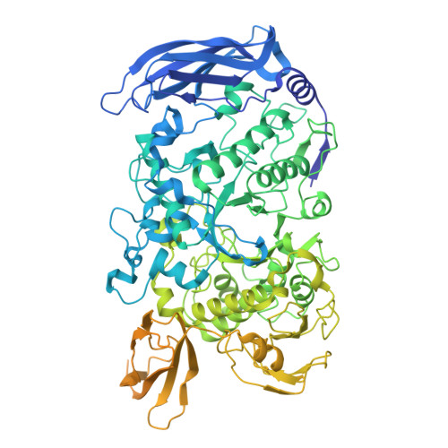
9QFA | pdb_00009qfa

Structure of the GH13 and MucBP domains of Ruminococcus bromii Amy12


Experimental Data Snapshot

  • Method: ELECTRON MICROSCOPY
  • Resolution: 3.10 Å
  • Aggregation State: PARTICLE 
  • Reconstruction Method: SINGLE PARTICLE 

Starting Model: experimental
View more details

wwPDB Validation   3D Report Full Report


This is version 1.0 of the entry. See complete history


Literature

Spatial and expression-driven regulation of the Ruminococcus bromii amylosome in resistant starch degradation

Wimmer, B.H.Morais, S.Amit, I.Tovar-Herrera, O.Tatli, M.Trautwein-Schult, A.Toedtli, P.Simoni, S.Lisibach, M.Levin, L.Koropatkin, N.Becher, D.Bayer, E.A.Medalia, O.Mizrahi, I.

To be published.

Macromolecules
Find similar proteins by:  (by identity cutoff)  |  3D Structure
Entity ID: 1
MoleculeChains Sequence LengthOrganismDetailsImage
Ruminococcus bromii Amy121,040Ruminococcus bromii L2-63Mutation(s): 0 
Gene Names: CBL15610.1
EC: 3.2.1.41
Entity Groups  
Sequence Clusters30% Identity50% Identity70% Identity90% Identity95% Identity100% Identity
Sequence Annotations
Expand
  • Reference Sequence
Experimental Data & Validation

Experimental Data

  • Method: ELECTRON MICROSCOPY
  • Resolution: 3.10 Å
  • Aggregation State: PARTICLE 
  • Reconstruction Method: SINGLE PARTICLE 
EM Software:
TaskSoftware PackageVersion
MODEL REFINEMENTPHENIX1.21_5207

Structure Validation

View Full Validation Report


Entry History & Funding Information

Deposition Data


Funding OrganizationLocationGrant Number
Swiss National Science FoundationSwitzerland10000885

Revision History  (Full details and data files)

  • Version 1.0: 2025-10-22
    Type: Initial release