9S6B | pdb_00009s6b

Aeropyrum pernix acylaminoacyl peptidase co-crystallized with meropenem.


Experimental Data Snapshot

  • Method: X-RAY DIFFRACTION
  • Resolution: 2.30 Å
  • R-Value Free: 
    0.229 (Depositor), 0.229 (DCC) 
  • R-Value Work: 
    0.197 (Depositor), 0.197 (DCC) 
  • R-Value Observed: 
    0.197 (Depositor) 

Starting Model: experimental
View more details

wwPDB Validation   3D Report Full Report


Ligand Structure Quality Assessment 


This is version 1.0 of the entry. See complete history


Literature

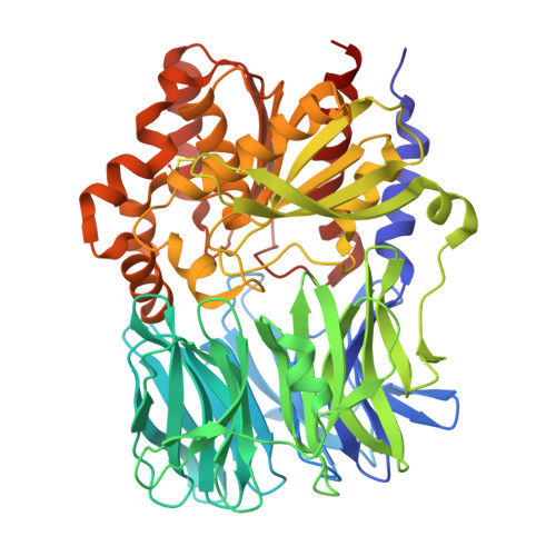
Ligand binding Pro-miscuity of acylpeptide hydrolase, structural analysis of a detoxifying serine hydrolase.

Kiss-Szeman, A.J.Takacs, L.Jakli, I.Banoczi, Z.Hosogi, N.Traore, D.A.K.Harmat, V.Perczel, A.Menyhard, D.K.

(2025) Protein Sci 34: e70320-e70320

  • DOI: https://doi.org/10.1002/pro.70320
  • Primary Citation of Related Structures:  
    9GNE, 9GOU, 9HXQ, 9S6B

  • PubMed Abstract: 

    Acylpeptide hydrolase (APEH) or acylaminoacyl-peptidase (AAP) is a serine hydrolase that regulates protein metabolism. It can also bind to and process unusual substrates, acting as a detoxifier. To better understand its promiscuous specificity, we determined the cryo-EM structures of mammalian APEH complexed with classical serine protease partners: a chloromethyl-ketone (CMK) inhibitor, an organophosphate (OP) pesticide (dichlorvos), and benzenesulfonyl-fluoride. Since CMK derivatives of N-acetylated peptides were suggested to induce apoptosis by inhibiting APEH, while OP complexes may serve as biomarkers of OP exposure and are linked to cognitive enhancement, these complexes carry physiological significance. We identified a unique strand-breaker Pro residue in the hydrolase domain, which relaxes the active site into a partially inactivated but more spacious conformation, transforming the classical serine protease apparatus into a versatile yet potent hydrolysis center with broad specificity, distinguishing the mammalian enzyme not only from other APEHs but also from serine α/β hydrolases sharing essentially the same fold.


  • Organizational Affiliation
    • Laboratory of Structural Chemistry and Biology, Institute of Chemistry, Eötvös Loránd University, Budapest, Hungary.

Macromolecules
Find similar proteins by:  (by identity cutoff)  |  3D Structure
Entity ID: 1
MoleculeChains Sequence LengthOrganismDetailsImage
Acylamino-acid-releasing enzyme
A, B, C, D
582Aeropyrum pernix K1Mutation(s): 1 
Gene Names: APE_1547.1
EC: 3.4.19.1
UniProt
Find proteins for Q9YBQ2 (Aeropyrum pernix (strain ATCC 700893 / DSM 11879 / JCM 9820 / NBRC 100138 / K1))
Explore Q9YBQ2 
Go to UniProtKB:  Q9YBQ2
Entity Groups  
Sequence Clusters30% Identity50% Identity70% Identity90% Identity95% Identity100% Identity
UniProt GroupQ9YBQ2
Sequence Annotations
Expand
  • Reference Sequence
Experimental Data & Validation

Experimental Data

  • Method: X-RAY DIFFRACTION
  • Resolution: 2.30 Å
  • R-Value Free:  0.229 (Depositor), 0.229 (DCC) 
  • R-Value Work:  0.197 (Depositor), 0.197 (DCC) 
  • R-Value Observed: 0.197 (Depositor) 
Space Group: P 1
Unit Cell:
Length ( Å )Angle ( ˚ )
a = 71.042α = 105.51
b = 98.283β = 103.19
c = 98.902γ = 100.39
Software Package:
Software NamePurpose
PHENIXrefinement
XDSdata reduction
XSCALEdata scaling
PHASERphasing

Structure Validation

View Full Validation Report


Ligand Structure Quality Assessment 


Entry History & Funding Information

Deposition Data


Funding OrganizationLocationGrant Number
European Regional Development FundEuropean UnionVEKOP-2.3.3-15-2017-00018
Hungarian National Research, Development and Innovation OfficeHungary2018-1.2.1-NKP-2018-00005
European Regional Development FundEuropean UnionRRF-2.3.1-21-2022-00015
Hungarian National Research, Development and Innovation OfficeHungary2020-1.1.6-JOVO-2021-00010

Revision History  (Full details and data files)

  • Version 1.0: 2025-10-22
    Type: Initial release