9DZN | pdb_00009dzn

KAT6A MYST domain complexed with a H3K14-CoA bisubstrate inhibitor


Experimental Data Snapshot

  • Method: X-RAY DIFFRACTION
  • Resolution: 1.72 Å
  • R-Value Free: 
    0.245 (Depositor), 0.242 (DCC) 
  • R-Value Work: 
    0.219 (Depositor), 0.217 (DCC) 
  • R-Value Observed: 
    0.220 (Depositor) 

Starting Model: experimental
View more details

wwPDB Validation   3D Report Full Report


Ligand Structure Quality Assessment 


This is version 1.1 of the entry. See complete history


Literature

Modulation of the substrate preference of a MYST acetyltransferase by a scaffold protein.

Sengupta, R.N.Brodsky, O.Bingham, P.Diehl, W.C.Ferre, R.Greasley, S.E.Johnson, E.Kraus, M.Lieberman, W.Meier, J.L.Paul, T.A.Maegley, K.A.

(2025) J Biological Chem 301: 108262-108262

  • DOI: https://doi.org/10.1016/j.jbc.2025.108262
  • Primary Citation of Related Structures:  
    9DZN

  • PubMed Abstract: 

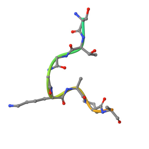
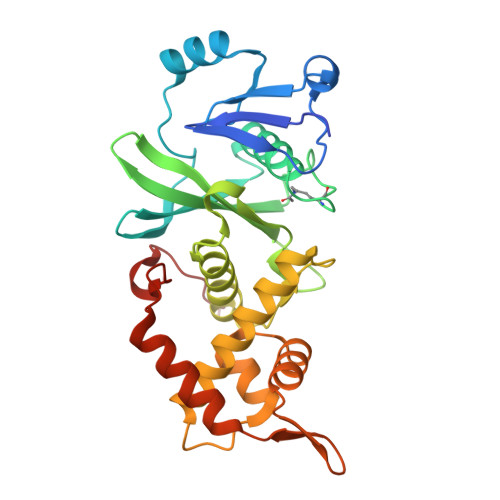
    The MYST family of lysine acetyltransferases are transcriptional regulators often dysregulated in cancer. In cells, MYST members form distinct multiprotein complexes that guide their histone substrate specificity, but how this selectivity is conferred is not fully understood. Here we interrogate a complex-mediated change in the substrate preference of the MYST member KAT6A, a target for cancer therapeutics. KAT6A forms a 4-protein complex with BRPF1, ING4/5, and MEAF6 to acetylate H3K23. However, additional substrates (H3K9, H3K14, and H3K27) have been proposed, and whether these residues are modified by KAT6A is unclear. We determined the histone substrate specificity of uncomplexed forms of KAT6A, including full-length KAT6A (KAT6A FL ) and the isolated acetyltransferase (MYST) domain, and the KAT6A FL 4-protein complex (KAT6A FL 4-plex). We show that the MYST domain and KAT6A FL preferentially acetylate H3K14, with this selectivity linked to a glycine pair preceding K14. A structure of the MYST domain bound to a H3K14-CoA bisubstrate inhibitor is consistent with a model in which the small size and flexibility of this glycine pair facilitates K14 acetylation. Notably, when KAT6A FL assembles into the 4-plex, H3K23 emerges as the favored substrate, with favorable recognition of an alanine-threonine pair before K23. These changes are mediated by BRPF1 and steady-state assays with H3 peptides indicate that this scaffold protein can alter the substrate preference of KAT6A FL by ≈10 3 -fold. Such context-dependent specificity illustrates how the functional properties of MYST members can be modulated by associated proteins and underscores the importance of characterizing these enzymes in their free and complexed forms.


  • Organizational Affiliation

    Oncology Research and Development, Pfizer, La Jolla, California, USA. Electronic address: raghuvir.sengupta@pfizer.com.


Macromolecules

Find similar proteins by:  Sequence   |   3D Structure  

Entity ID: 1
MoleculeChains Sequence LengthOrganismDetailsImage
Histone H3K14A [auth C]19Homo sapiensMutation(s): 0 
UniProt & NIH Common Fund Data Resources
Find proteins for Q6NXT2 (Homo sapiens)
Explore Q6NXT2 
Go to UniProtKB:  Q6NXT2
PHAROS:  Q6NXT2
GTEx:  ENSG00000188375 
Entity Groups  
Sequence Clusters30% Identity50% Identity70% Identity90% Identity95% Identity100% Identity
UniProt GroupQ6NXT2
Sequence Annotations
Expand
  • Reference Sequence
Find similar proteins by:  (by identity cutoff)  |  3D Structure
Entity ID: 2
MoleculeChains Sequence LengthOrganismDetailsImage
Histone acetyltransferase KAT6AB [auth A]286Homo sapiensMutation(s): 0 
Gene Names: KAT6AMOZMYST3RUNXBP2ZNF220
EC: 2.3.1.48
UniProt & NIH Common Fund Data Resources
Find proteins for Q92794 (Homo sapiens)
Explore Q92794 
Go to UniProtKB:  Q92794
PHAROS:  Q92794
GTEx:  ENSG00000083168 
Entity Groups  
Sequence Clusters30% Identity50% Identity70% Identity90% Identity95% Identity100% Identity
UniProt GroupQ92794
Sequence Annotations
Expand
  • Reference Sequence
Small Molecules
Experimental Data & Validation

Experimental Data

  • Method: X-RAY DIFFRACTION
  • Resolution: 1.72 Å
  • R-Value Free:  0.245 (Depositor), 0.242 (DCC) 
  • R-Value Work:  0.219 (Depositor), 0.217 (DCC) 
  • R-Value Observed: 0.220 (Depositor) 
Space Group: P 41 21 2
Unit Cell:
Length ( Å )Angle ( ˚ )
a = 59.368α = 90
b = 59.368β = 90
c = 210.05γ = 90
Software Package:
Software NamePurpose
BUSTERrefinement
XDSdata reduction
STARANISOdata scaling
BUSTERphasing

Structure Validation

View Full Validation Report


Ligand Structure Quality Assessment 


Entry History & Funding Information

Deposition Data


Funding OrganizationLocationGrant Number
Not fundedUnited States--

Revision History  (Full details and data files)

  • Version 1.0: 2025-02-19
    Type: Initial release
  • Version 1.1: 2025-03-26
    Changes: Database references