9LSI | pdb_00009lsi

Cryo-EM structure of the S82C-S82C diabody complex (CitS-diabody #2-TLR3)


Experimental Data Snapshot

  • Method: ELECTRON MICROSCOPY
  • Resolution: 3.30 Å
  • Aggregation State: PARTICLE 
  • Reconstruction Method: SINGLE PARTICLE 

wwPDB Validation   3D Report Full Report


This is version 1.2 of the entry. See complete history


Literature

Disulfide-stabilized diabodies enable near-atomic cryo-EM imaging of small proteins: A case study of the bacterial Na + /citrate symporter CitS.

Kim, S.Kim, J.W.Park, J.G.Lee, S.S.Choi, S.H.Lee, J.O.Jin, M.S.

(2025) Structure 33: 1088-1100.e3

  • DOI: https://doi.org/10.1016/j.str.2025.03.006
  • Primary Citation of Related Structures:  
    9LSH, 9LSI, 9LSJ, 9LSK

  • PubMed Abstract: 

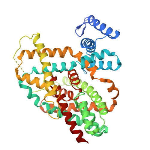
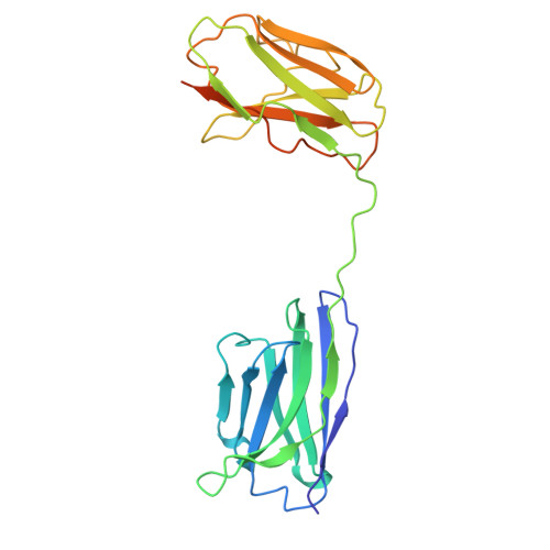
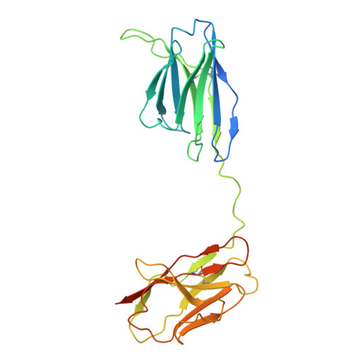
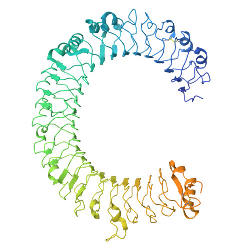
    Diabodies are engineered antibody fragments with two antigen-binding Fv domains. Previously, we demonstrated that they are often highly flexible but can be rigidified by introducing a disulfide bond at the Fv interface. In this study, we explored the potential of disulfide-bridged, bispecific diabodies for near-atomic cryoelectron microscopy (cryo-EM) imaging of small proteins because they can predictably link target proteins to "structural marker" proteins. As a case study, we used the bacterial citrate transporter CitS as the target protein, and the horseshoe-shaped ectodomain of human Toll-like receptor 3 (TLR3) as the marker. We show that diabodies containing one or two disulfide bonds enabled the 3D reconstruction of CitS at resolutions of 3.3 Å and 3.1 Å, respectively. This resolution surpassed previous crystallographic results and allowed us to visualize the high-resolution structural features of the transporter. Our work expands the application of diabodies in structural biology to address a key limitation in the field.


  • Organizational Affiliation
    • Department of Life Sciences, Gwangju Institute of Science and Technology, Gwangju 61005, Republic of Korea.

Macromolecules
Find similar proteins by:  (by identity cutoff)  |  3D Structure
Entity ID: 1
MoleculeChains Sequence LengthOrganismDetailsImage
Citrate/sodium symporter
A, B
446Klebsiella pneumoniaeMutation(s): 0 
Gene Names: citS
Membrane Entity: Yes 
UniProt
Find proteins for P31602 (Klebsiella pneumoniae)
Explore P31602 
Go to UniProtKB:  P31602
Entity Groups  
Sequence Clusters30% Identity50% Identity70% Identity90% Identity95% Identity100% Identity
UniProt GroupP31602
Sequence Annotations
Expand
  • Reference Sequence
Find similar proteins by:  (by identity cutoff)  |  3D Structure
Entity ID: 2
MoleculeChains Sequence LengthOrganismDetailsImage
Diabody (CitS VH-TLR3 VL)256synthetic constructMutation(s): 0 
Entity Groups  
Sequence Clusters30% Identity50% Identity70% Identity90% Identity95% Identity100% Identity
Sequence Annotations
Expand
  • Reference Sequence
Find similar proteins by:  (by identity cutoff)  |  3D Structure
Entity ID: 3
MoleculeChains Sequence LengthOrganismDetailsImage
Diabody (TLR3 VH-CitS VL)266synthetic constructMutation(s): 0 
Entity Groups  
Sequence Clusters30% Identity50% Identity70% Identity90% Identity95% Identity100% Identity
Sequence Annotations
Expand
  • Reference Sequence
Find similar proteins by:  (by identity cutoff)  |  3D Structure
Entity ID: 4
MoleculeChains Sequence LengthOrganismDetailsImage
Toll-like receptor 3904Homo sapiensMutation(s): 0 
Gene Names: TLR3
Membrane Entity: Yes 
UniProt & NIH Common Fund Data Resources
Find proteins for O15455 (Homo sapiens)
Explore O15455 
Go to UniProtKB:  O15455
PHAROS:  O15455
GTEx:  ENSG00000164342 
Entity Groups  
Sequence Clusters30% Identity50% Identity70% Identity90% Identity95% Identity100% Identity
UniProt GroupO15455
Glycosylation
Glycosylation Sites: 9Go to GlyGen: O15455-1
Sequence Annotations
Expand
  • Reference Sequence
Oligosaccharides

Help

Entity ID: 5
MoleculeChains Length2D Diagram Glycosylation3D Interactions
2-acetamido-2-deoxy-beta-D-glucopyranose-(1-4)-2-acetamido-2-deoxy-beta-D-glucopyranoseF,
G,
I [auth J]
2N-Glycosylation
Glycosylation Resources
GlyTouCan:  G42666HT
GlyCosmos:  G42666HT
GlyGen:  G42666HT
Entity ID: 6
MoleculeChains Length2D Diagram Glycosylation3D Interactions
2-acetamido-2-deoxy-beta-D-glucopyranose-(1-4)-2-acetamido-2-deoxy-beta-D-glucopyranose-(1-4)-2-acetamido-2-deoxy-beta-D-glucopyranoseH [auth I]3N-Glycosylation
Glycosylation Resources
GlyTouCan:  G47362BJ
GlyCosmos:  G47362BJ
GlyGen:  G47362BJ
Small Molecules
Ligands 3 Unique
IDChains Name / Formula / InChI Key2D Diagram3D Interactions
PLM
Query on PLM

Download Ideal Coordinates CCD File 
K [auth A]PALMITIC ACID
C16 H32 O2
IPCSVZSSVZVIGE-UHFFFAOYSA-N
NAG
Query on NAG

Download Ideal Coordinates CCD File 
L [auth E],
M [auth E],
N [auth E],
O [auth E],
P [auth E]
2-acetamido-2-deoxy-beta-D-glucopyranose
C8 H15 N O6
OVRNDRQMDRJTHS-FMDGEEDCSA-N
CIT (Subject of Investigation/LOI)
Query on CIT

Download Ideal Coordinates CCD File 
J [auth A]CITRIC ACID
C6 H8 O7
KRKNYBCHXYNGOX-UHFFFAOYSA-N
Experimental Data & Validation

Experimental Data

  • Method: ELECTRON MICROSCOPY
  • Resolution: 3.30 Å
  • Aggregation State: PARTICLE 
  • Reconstruction Method: SINGLE PARTICLE 
EM Software:
TaskSoftware PackageVersion
MODEL REFINEMENTPHENIX1.18.2_3874

Structure Validation

View Full Validation Report


Entry History & Funding Information

Deposition Data


Funding OrganizationLocationGrant Number
Samsung Science and Technology FoundationKorea, Republic OfSSTF-BA1702-14
National Research Foundation (NRF, Korea)Korea, Republic OfRS-2024-00344154
National Research Foundation (NRF, Korea)Korea, Republic OfRS-2024-00440614

Revision History  (Full details and data files)

  • Version 1.0: 2025-03-12
    Type: Initial release
  • Version 1.1: 2025-04-16
    Changes: Data collection, Database references
  • Version 1.2: 2025-06-18
    Changes: Data collection, Database references