9LVC | pdb_00009lvc

Temperature induces a shift from the dihexamer to the hexamer form of insulin


Experimental Data Snapshot

  • Method: X-RAY DIFFRACTION
  • Resolution: 2.30 Å
  • R-Value Free: 
    0.287 (Depositor), 0.289 (DCC) 
  • R-Value Work: 
    0.232 (Depositor), 0.234 (DCC) 
  • R-Value Observed: 
    0.237 (Depositor) 

Starting Model: experimental
View more details

wwPDB Validation   3D Report Full Report


Ligand Structure Quality Assessment 


This is version 1.1 of the entry. See complete history


Literature

Temperature induces a shift from insulin dihexamer to hexamer in collective dynamics.

Ayan, E.

(2025) Protein Sci 34: e70245-e70245

  • DOI: https://doi.org/10.1002/pro.70245
  • Primary Citation of Related Structures:  
    9LVC, 9LVD, 9LVE

  • PubMed Abstract: 

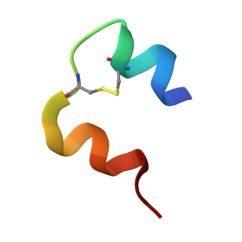
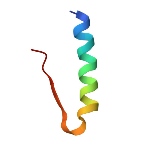
    Structures based on x-ray diffraction data collected at 2.3, 2.88, and 2.95 Å resolutions have been determined for long-acting dihexamer insulin at three different temperatures, ranging from 100 to 300 K. It has been observed that the unit-cell parameters of the insulin crystal at 100 K change at 200 K. This change is likely due to the subtle repacking of the rhombohedral insulin crystal and the loss of noncovalent interactions involving myristic acid, which binds two hexamers. Computational analyses indicate that allosteric residues and fatty acid-binding residues of insulin hexamers exhibit reduced collective dynamics and inter-residue coupling, possibly resulting from increased structural fluctuations due to elevated thermal vibrations. This transition has been observed at a characteristic temperature of 200 K, potentially highlighting underlying alterations in the dynamic structure of the fatty acid-solvent interface in the dimer of hexamers. Combined with computational analyses, these findings provide key insights into thermal stability mechanisms, which are crucial for developing thermostable insulin formulations in industrial applications.


  • Organizational Affiliation
    • Center for Targeted Therapy Center, Bogazici University, Istanbul, Turkey.

Macromolecules

Find similar proteins by:  Sequence   |   3D Structure  

Entity ID: 1
MoleculeChains Sequence LengthOrganismDetailsImage
Insulin A chain
A, C, E, G
21Homo sapiensMutation(s): 0 
Gene Names: INS
UniProt & NIH Common Fund Data Resources
Find proteins for P01308 (Homo sapiens)
Explore P01308 
Go to UniProtKB:  P01308
PHAROS:  P01308
GTEx:  ENSG00000254647 
Entity Groups  
Sequence Clusters30% Identity50% Identity70% Identity90% Identity95% Identity100% Identity
UniProt GroupP01308
Sequence Annotations
Expand
  • Reference Sequence
Find similar proteins by:  (by identity cutoff)  |  3D Structure
Entity ID: 2
MoleculeChains Sequence LengthOrganismDetailsImage
Insulin B chain
B, D, F, H
29Homo sapiensMutation(s): 0 
Gene Names: INS
UniProt & NIH Common Fund Data Resources
Find proteins for P01308 (Homo sapiens)
Explore P01308 
Go to UniProtKB:  P01308
PHAROS:  P01308
GTEx:  ENSG00000254647 
Entity Groups  
Sequence Clusters30% Identity50% Identity70% Identity90% Identity95% Identity100% Identity
UniProt GroupP01308
Sequence Annotations
Expand
  • Reference Sequence
Small Molecules
Ligands 4 Unique
IDChains Name / Formula / InChI Key2D Diagram3D Interactions
MYR (Subject of Investigation/LOI)
Query on MYR

Download Ideal Coordinates CCD File 
L [auth B],
P [auth D],
T [auth F],
X [auth H]
MYRISTIC ACID
C14 H28 O2
TUNFSRHWOTWDNC-UHFFFAOYSA-N
IPH (Subject of Investigation/LOI)
Query on IPH

Download Ideal Coordinates CCD File 
I [auth B],
M [auth D],
Q [auth F],
U [auth H]
PHENOL
C6 H6 O
ISWSIDIOOBJBQZ-UHFFFAOYSA-N
ZN (Subject of Investigation/LOI)
Query on ZN

Download Ideal Coordinates CCD File 
J [auth B],
N [auth D],
R [auth F],
V [auth H]
ZINC ION
Zn
PTFCDOFLOPIGGS-UHFFFAOYSA-N
CL (Subject of Investigation/LOI)
Query on CL

Download Ideal Coordinates CCD File 
K [auth B],
O [auth D],
S [auth F],
W [auth H]
CHLORIDE ION
Cl
VEXZGXHMUGYJMC-UHFFFAOYSA-M
Experimental Data & Validation

Experimental Data

  • Method: X-RAY DIFFRACTION
  • Resolution: 2.30 Å
  • R-Value Free:  0.287 (Depositor), 0.289 (DCC) 
  • R-Value Work:  0.232 (Depositor), 0.234 (DCC) 
  • R-Value Observed: 0.237 (Depositor) 
Space Group: H 3
Unit Cell:
Length ( Å )Angle ( ˚ )
a = 79.064α = 90
b = 79.064β = 90
c = 79.496γ = 120
Software Package:
Software NamePurpose
PHENIXrefinement
HKL-2000data reduction
CrysalisProdata scaling
PHENIXphasing

Structure Validation

View Full Validation Report


Ligand Structure Quality Assessment 


Entry History & Funding Information

Deposition Data


Funding OrganizationLocationGrant Number
Other government--

Revision History  (Full details and data files)

  • Version 1.0: 2025-02-26
    Type: Initial release
  • Version 1.1: 2025-09-10
    Changes: Database references