9O4F | pdb_00009o4f

Pre-fusion Stabilized HERV-K Envelope Trimer Ectodomain


Experimental Data Snapshot

  • Method: ELECTRON MICROSCOPY
  • Resolution: 2.24 Å
  • Aggregation State: PARTICLE 
  • Reconstruction Method: SINGLE PARTICLE 

Starting Model: in silico
View more details

wwPDB Validation   3D Report Full Report


This is version 1.1 of the entry. See complete history


Literature

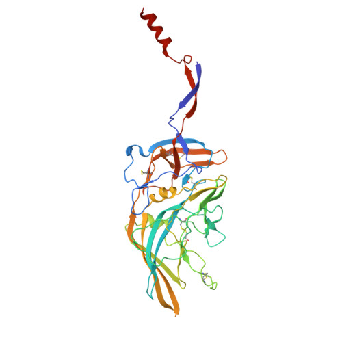
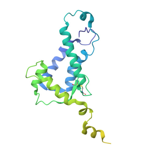
Human endogenous retrovirus K (HERV-K) envelope structures in pre- and postfusion by cryo-EM.

Shek, J.Sun, C.Wilson, E.M.Moadab, F.Hastie, K.M.Rajamanickam, R.R.Penalosa, P.J.Harkins, S.S.Parekh, D.Hariharan, C.Zyla, D.S.Yu, C.Shaffer, K.C.L.Lewis, V.I.Avalos, R.D.Mustelin, T.Saphire, E.O.

(2025) Sci Adv 11: eady8168-eady8168

  • DOI: https://doi.org/10.1126/sciadv.ady8168
  • Primary Citation of Related Structures:  
    9MLA, 9MLK, 9O4F

  • PubMed Abstract: 

    Human endogenous retroviruses (HERVs) are remnants of ancient infections that comprise ~8% of the human genome. The HERV-K envelope glycoprotein (Env) is aberrantly expressed in cancers, autoimmune disorders, and neurodegenerative diseases, and is targeted by patients' own antibodies. However, a lack of structural information has limited molecular and immunological studies of the roles of HERVs in disease. Here, we present cryo-electron microscopy structures of stabilized HERV-K Env in the prefusion conformation, revealing a distinct fold and architecture compared to HIV and simian immunodeficiency virus. We also generated and characterized a panel of monoclonal antibodies with subunit and conformational specificity, serving as valuable research tools. These antibodies enabled structure determination of the postfusion conformation of HERV-K Env, including its unique "tether" helix, and antibody-bound prefusion Env. Together, these results provide a structural framework that opens the door to mechanistic studies of HERV-K Env and tools for its evaluation as a potential therapeutic target.


  • Organizational Affiliation
    • Center for Vaccine Innovation, La Jolla Institute for Immunology, La Jolla, CA 92037, USA.

Macromolecules
Find similar proteins by:  (by identity cutoff)  |  3D Structure
Entity ID: 1
MoleculeChains Sequence LengthOrganismDetailsImage
Surface protein
A, B, C
369Homo sapiensMutation(s): 3 
Gene Names: ERVK-6ERVK6
UniProt
Find proteins for Q69384 (Homo sapiens)
Explore Q69384 
Go to UniProtKB:  Q69384
Entity Groups  
Sequence Clusters30% Identity50% Identity70% Identity90% Identity95% Identity100% Identity
UniProt GroupQ69384
Glycosylation
Glycosylation Sites: 6
Sequence Annotations
Expand
  • Reference Sequence
Find similar proteins by:  (by identity cutoff)  |  3D Structure
Entity ID: 2
MoleculeChains Sequence LengthOrganismDetailsImage
Transmembrane protein,Fibritin
D, E, F
253Homo sapiensTequatrovirus T4
This entity is chimeric
Mutation(s): 1 
Gene Names: ERVK-6ERVK6
UniProt
Find proteins for P10104 (Enterobacteria phage T4)
Explore P10104 
Go to UniProtKB:  P10104
Find proteins for Q69384 (Homo sapiens)
Explore Q69384 
Go to UniProtKB:  Q69384
Entity Groups  
Sequence Clusters30% Identity50% Identity70% Identity90% Identity95% Identity100% Identity
UniProt GroupsQ69384P10104
Glycosylation
Glycosylation Sites: 4
Sequence Annotations
Expand
  • Reference Sequence
Oligosaccharides

Help

Entity ID: 3
MoleculeChains Length2D Diagram Glycosylation3D Interactions
alpha-D-mannopyranose-(1-3)-[alpha-D-mannopyranose-(1-6)]beta-D-mannopyranose-(1-4)-2-acetamido-2-deoxy-beta-D-glucopyranose-(1-4)-2-acetamido-2-deoxy-beta-D-glucopyranose
G, L, Q
5N-Glycosylation
Glycosylation Resources
GlyTouCan:  G22768VO
GlyCosmos:  G22768VO
GlyGen:  G22768VO
Entity ID: 4
MoleculeChains Length2D Diagram Glycosylation3D Interactions
2-acetamido-2-deoxy-beta-D-glucopyranose-(1-4)-2-acetamido-2-deoxy-beta-D-glucopyranose
H, J, K, M, O
H, J, K, M, O, P, R, T, U
2N-Glycosylation
Glycosylation Resources
GlyTouCan:  G42666HT
GlyCosmos:  G42666HT
GlyGen:  G42666HT
Entity ID: 5
MoleculeChains Length2D Diagram Glycosylation3D Interactions
beta-D-mannopyranose-(1-4)-2-acetamido-2-deoxy-beta-D-glucopyranose-(1-4)-2-acetamido-2-deoxy-beta-D-glucopyranose
BA [auth b],
CA [auth c],
I,
N,
S,
BA [auth b],
CA [auth c],
I,
N,
S,
V,
W,
Y,
Z
3N-Glycosylation
Glycosylation Resources
GlyTouCan:  G15407YE
GlyCosmos:  G15407YE
GlyGen:  G15407YE
Entity ID: 6
MoleculeChains Length2D Diagram Glycosylation3D Interactions
beta-D-mannopyranose-(1-4)-2-acetamido-2-deoxy-beta-D-glucopyranose-(1-4)-[alpha-L-fucopyranose-(1-6)]2-acetamido-2-deoxy-beta-D-glucopyranoseAA [auth a],
DA [auth d],
X
4N-Glycosylation
Glycosylation Resources
GlyTouCan:  G32152BH
GlyCosmos:  G32152BH
GlyGen:  G32152BH
Experimental Data & Validation

Experimental Data

  • Method: ELECTRON MICROSCOPY
  • Resolution: 2.24 Å
  • Aggregation State: PARTICLE 
  • Reconstruction Method: SINGLE PARTICLE 
EM Software:
TaskSoftware PackageVersion
MODEL REFINEMENTPHENIX1.21.1_5286
RECONSTRUCTIONcryoSPARC4.6.0

Structure Validation

View Full Validation Report


Entry History & Funding Information

Deposition Data


Funding OrganizationLocationGrant Number
Other privateUnited States18030-01-000-408
Other privateUnited States13502-01-000-408

Revision History  (Full details and data files)

  • Version 1.0: 2025-07-30
    Type: Initial release
  • Version 1.1: 2025-09-03
    Changes: Data collection, Database references